


MedFriendly®


Anterolisthesis
To understand what anterolisthesis means it is first
necessary to understand the meaning of the word
vertebrae. Vertebrae are bones that form an opening in
which the spinal cord passes. These bones are stacked
one on top of another, as shown in the picture below.
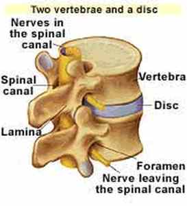
Each individual bone that makes up the vertebrae is
called a vertebra. The thick, drum-shaped part of the
bone that forms the front part of the vertebra is known as
the vertebral body. In the picture above, the vertebral
bodies are located in the front part of the picture,
between the red areas. The red areas are discs.
Because these discs are positioned between the
vertebrae, they are known as interverterbral discs. The
discs are flat and cushiony and act as shock absorbers.
Normal appearing vertebrae.
FEATURED BOOK: Clinical Anatomy of the Spine
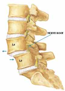
In anterolisthesis, the upper vertebral body is positioned abnormally compared to the
lower vertebral body. More specifically, the upper vertebral body slips forward upon the
one below it. The amount of slippage is graded on a scale from 1 to 4 (or I to IV in
Roman numerals). Each grade (starting at grade I) means that the slippage has
increased another 25% of the vertebral body. So Grade I anterolisthesis means that
there has been 25% slippage, Grade II antherolisthesis means there has been 50%
slippage, Grade III anterolisthesis means there has been 75% slippage, and Grade IV
anterolisthesis means there has been 100% slippage. Thus, Grade I anterolisthesis is
mild and Grade IV anterolisthesis is severe. Anterolisthesis is often caused by bone
fractures.
"Where Medical Information is Easy to Understand"™
WHAT DOES ANTEROLISTHESIS LOOK LIKE?
A picture of normal appearing vertebrae appears above. Below it a
picture of anterolisthesis in the 4th lumbar vertebrae on the 5th
lumbar vertebrae.
HOW IS ANTEROLISTHESIS TREATED?
If the condition is bad enough and has not responded to
conservative treatment such as rest and physical therapy,
anterolisthesis is treated through a surgical technique known as
interbody fusion.
In this technique, an incision is made in the back, through the middle layer of muscles and ligaments
that sit on either side of the spine. A ligament is a tough band of tissue that attaches to joints and holds
them together. A joint is a place where two bones contact each other.
Cutting into the above areas frees the attachments to the spinous processes
and laminae. Spinous processes are the bony parts that project out from the
back of the vertebrae. Some of the back muscles are attached to the spinous
processes. The laminae are the thin, flattened part of the vertebral arch. The
vertebral arch is the ring of bone that, together with the vertebral bodies,
surrounds the spinal cord. A picture of a lamina is included below. The areas
projecting outwards are the spinous processes.
A small instrument is then used which removes small bits of bone from the
lamina until the nerves can be seen. The nerves are moved slightly to expose
the intervertebral discs (see above). Using various instruments, the disc is
removed through the right and left side of the spinal canal. The spinal canal is
the space between the spinal cord and the bony structure that surrounds it.
Once the disc is removed, the doctor aligns the vertebrae. The empty space where the disc used to be
is then filled with bone. The bone is taken from other parts of the body. In some cases, the space is
filled with artificial bone, which is known as a bone block. With the space between the vertebrae being
filled, the vertebrae are fused together. Thus, the vertebrae can no longer slip forward on the one below
it. The fused vertebrae are often stabilized further with hardware such as screws.
More recently, doctors have been implanting a device known as a spinal cage in the space between the
vertebrae. A spinal cage is literally a small threaded cage that is filled with small pieces of natural bone.
The packed cage is then capped and implanted between the vertebrae. As natural bone grows through
holes in the cage to fuse with the natural bone inside it, permanent fusion and stability is accomplished.
WHAT IS THE ORIGIN OF THE TERM, ANTEROLISTHESIS?
Anterolisthesis comes from the Latin word "ante" meaning "front," and the Greek word "listhesis"
meaning "to slide down a slippery path." Put the words together and you have "to slide down a slippery
front(wards) path.