


MedFriendly®


Cerebral Aqueduct
The cerebral aqueduct is a narrow opening
in the brain that connects the third ventricle
with the fourth ventricle, allowing
cerebrospinal fluid to flow between these
two areas. Ventricles are openings in the
brain that provide a pathway for
cerebrospinal fluid.
Cerebrospinal fluid is the cushiony fluid that
protects the brain and spine from trauma.
The cerebral aqueduct drains cerebrospinal
fluid from the third ventricle to the fourth
ventricle.
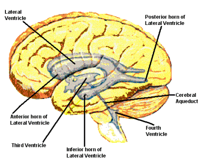
The third ventricle is a narrow, four-sided, irregularly shaped opening in the middle of the
brain. The fourth ventricle is a wide, flat open space located in the back bottom part of
the brain. The third ventricle, fourth ventricle, and cerebral aqueduct are pictured below.
FEATURED BOOK: Neuroanatomy Through Clinical Cases (2nd ed.)
WHAT ARE SOME OTHER DETAILS ABOUT THE CEREBRAL AQUEDUCT?
The cerebral aqueduct is about 3/4 of an inch long and is lined with ependyma.
Ependyma is a type of membrane that lines the ventricles of the brain.
"Where Medical Information is Easy to Understand"™
A membrane is a thin layer of flexible tissue that covers something.
The cerebral aqueduct can be found in the midbrain. The midbrain
is the top part of the brainstem. The brainstem is an area in the
lower part of the brain that connects it with the spinal cord. Below
is a picture of the midbrain and the rest of the brainstem (pons and
medulla).
The cerebral aqueduct is the only major part of the ventricle system
that does not contain a choroid plexus. The choroid plexus is a
cluster of blood vessels that lines the ventricles and produce
cerebrospinal fluid.
WHAT HAPPENS IF THE CEREBRAL AQUEDUCT BECOMES BLOCKED?
The cerebral aqueduct can become blocked. This leads to an abnormal increase in cerebrospinal fluid in
the brain and enlargement of the ventricles, a condition known as hydrocephalus. Cerebrospinal fluid is
the cushiony fluid that protects the brain and spine from trauma. The cerebrospinal fluid builds up when
there is a blockage because there is no place for it to go, much like the buildup of fluid that occurs when
a pipe in the sink becomes clogged. The ventricles become larger because the increased fluid (which
has no place to go) builds up and expands outwards.
In the case of a blockage of the cerebral aqueduct, cerebrospinal fluid from the lateral (side) ventricles
and the third ventricle cannot get down to the fourth ventricle. The lateral ventricles are two curved
openings (shaped like a horseshoe) located deep within the top section of the brain. There is one lateral
ventricle on each side of the brain. See above for a picture of the lateral ventricles.
WHAT ELSE IS THE CEREBRAL AQUEDUCT KNOWN AS?
The lateral ventricle is also known as the aqueduct of sylvius, aqueductus sylvii, sylvian aqueduct,
aqueductus mesencephali, aqueductus cerebri, aqueduct of cerebrum, and iter a tertio ad quartum
ventriculum.
WHAT IS THE ORIGIN OF THE TERM, CEREBRAL AQUEDUCT?
Cerebral aqueduct comes from the Latin word "cerebrum" meaning "brain" and the Latin word
"aqueductus" meaning "water canal." Put the words together and you have "brain water canal."