MedFriendly®


Echogenic Cardiac Focus
Echogenic cardiac foci refer to a high number of
echoes inside an area (see next section) of an unborn
childs heart. The high number of echoes shows up as
bright spots (that resemble a small white pea or pearl)
in the heart on an echocardiography. Echocardiography
is an imaging technique that uses types of sound
waves to produce pictures of the heart. The word foci
in echogenic cardiac foci refers to more than one
area in the heart where there are abnormally high
numbers of echoes. The word focus in echogenic
cardiac focus refers to a single area in the heart where
there are abnormally high numbers of echoes.
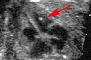
The arrow shows an echogenic
cardiac focus.
FEATURED BOOK: Clinical Cardiology Made Ridiculously Simple
WHERE ARE ECHOGENIC CARDIAC FOCI LOCATED?
Most echogenic cardiac foci are located in an area of the heart known as the left
ventricle. The left ventricle is an opening in the heart that pumps out blood with a high
level of oxygen to the body. However, echogenic cardiac foci can also be found in the
right ventricle. The right ventricle is an opening in the heart that pumps out blood with the
low level of oxygen to the lungs.
ARE ECHOGENIC CARDIAC FOCI UNUSUAL OR ABNORMAL?
No. Echogenic cardiac foci are not an unusual or abnormal finding in an
echocardiography of the heart.
"Where Medical Information is Easy to Understand"™
In fact, up to 7.4% of unborn children have echogenic cardiac foci
when the mother is in the second trimester (months 3 to 6). The
vast majority of children with echogenic cardiac foci are born
normal. This is especially the case for the children of mothers who
are under 35 years of age. Only rarely are echogenic cardiac foci
associated with birth defects. It has been suggested that the
presence of echogenic cardiac foci is associated with a greater risk
of the child having Down syndrome. Down syndrome is an
abnormality that is present from birth that results in mental
impairments and a characteristic physical appearance (small facial
features, large tongue that sticks out, a flat back area of the head,
and hands that are short and broad).
There is conflicting evidence, however, as to whether echogenic foci in the heart lead to an increased risk
for Down syndrome. That is, some studies have found that there is no increased risk whereas others
have found that unborn children with echogenic cardiac foci are four times more likely to have Down
syndrome. Many studies suggest that unborn children with echogenic cardiac foci have about a 1%
chance of having Down syndrome.
WHAT CAUSES ECHOGENIC CARDIAC FOCI?
The formation of calcium or other minerals in the ventricles is what is thought to be the cause of the
increased echoing that leads to echogenic cardiac foci. Calcium is a natural element that is very important
in bone formation. As evidence of this, a study of the heart tissue of unborn children with Down syndrome
found a significantly increased calcification (formation of calcium) in the papillary muscle of the heart. The
papillary muscle of the heart is a rounded or cone-shaped type of muscle found in the ventricles of the
heart.
DO ECHOGENIC CARDIAC FOCI GO AWAY?
Yes. Many of the cases of echogenic cardiac foci go away by themselves by the time the child is born. In
more than half of the cases, echogenic cardiac foci go away by age 5.
WHAT SHOULD PARENTS DO IF THE UNBORN CHILD HAS ECHOGENIC CARDIAC FOCI?
If the only finding is echogenic cardiac foci, doctors usually will not do additional testing unless there are
other abnormalities present and/or additional reasons to be concerned about the structure and functioning
of the heart. Doctors usually recommend that the mother have more detailed ultrasound testing done to
see if there are any additional signs that the child has Down syndrome. Ultrasound testing uses certain
types of sound waves to produce images of the body (in this case, the unborn child).
Amniocentesis is usually not recommended if echogenic cardiac foci are the only finding. Amniocentesis is
a procedure in which a small amount of fluid (known as amniotic fluid) is withdrawn from the sac that
surrounds the unborn child in the mothers uterus. This fluid can then be tested to detect abnormalities in
the unborn child. The uterus is a hollow organ in a female's body where the egg is implanted and the baby
develops. Doctors may recommend amniocentesis, however, if the mother is 35 years or older, if there
are additional abnormal results on the echocardiography, or if there are abnormal blood test results.
HOW LONG HAVE DOCTORS KNOWN ABOUT ECHOGENIC CARDIAC FOCI?
Echogenic cardiac foci were first described in the mid-1980s.
WHAT ELSE ARE ECHOGENIC CARDIAC FOCI KNOWN AS?
Echogenic cardiac foci are also known as cardiac echogenic foci/focus, intracardiac echogenic
foci/focus, echogenic foci/focus of the heart, and echogenic intracardiac foci/focus.
WHAT IS THE ORIGIN OF THE TERM, ECHOGENIC CARDIAC FOCI?
Echogenic cardiac foci comes from the Greek word "echo" meaning "sound," the Greek word genos
meaning birth, the Greek word "ikos" meaning "pertaining to," the Greek word "kardia" meaning "heart,"
and the Latin word focus meaning hearth. Put the words together and you have "pertaining to (the) birth
(of) sound (in the) heart hearth."