


MedFriendly®


Lacrimal Apparatus
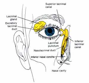
The lacrimal apparatus (pictured to the right) is the
system in the body that produces and drains tears. The
lacrimal apparatus is made up of many parts, described
below.
WHAT PARTS MAKE UP THE LACRIMAL
APPARATUS?
The lacrimal apparatus is comprised of many parts.
Here is a brief description.
LACRIMAL GLANDS: Small organs that excrete
(release) tears.
FEATURED BOOK: The Dry Eye Remedy
There are two types of lacrimal glands, known as main and accessory lacrimal glands.
The main lacrimal glands (located in the upper and outer part of the eye sockets) release
extra tears, such as when the eye is irritated and during crying. The main lacrimal glands
drain tears into the conjunctiva. The conjunctiva is a layer that covers and protects the
inside of the eyelids and the front part of the sclera (the white part of the eyes). The
accessory lacrimal glands (located within the conjunctiva) maintain a normal amount of
tears on the surface of the conjunctiva. This helps overcome the effect of tears
evaporating (changing from a liquid to a gas).
"Where Medical Information is Easy to Understand"™
LACRIMAL LAKE: A small open area of the conjunctiva of the eye
where tears collect after bathing in the front part of the eyeball and
in a slit-like space known as the conjunctival sac. The conjunctival
sac is between the eyelids and the conjunctiva-covered eyeball.
EXCRETORY LACRIMAL DUCTS: Tube shaped areas that release
tears from the lacrimal gland into the top part of the conjunctiva sac.
LACRIMAL PUNCTA: Tiny openings towards the inner part of each
eyelid, that tears drain through. Picture the lacrimal puncta as being
like a drain in a kitchen sink. The lacrimal puncta connect and send
tears to narrow tubes, known as lacrimal canals.
LACRIMAL CANALS (SUPERIOR & INFERIOR): Curved, tube shaped structures connected to the
lacrimal puncta, that tears coming from the lacrimal lake drain into. As you can see in the picture, the
superior (above) lacrimal canal is on the top and the inferior (below) lacrimal canal is on the bottom. Tears
travel from the lacrimal canals to the lacrimal sacs. The lacrimal canals are also known as the lacrimal
ducts and lacrimal canaliculi.
LACRIMAL SACS: Hollow spaces that the lacrimal canals (see above) drain tears into. Each eye has a
lacrimal sac for tears to drain into. The yellow structure pictured above, next to the lacrimal canals, is the
lacrimal sac. Flat muscles that cover the lacrimal sac, squeeze and release it during blinking. This helps
produce a suction effect that draws away extra tears when blinking. This is why people blink when they
cry.
LACRIMAL BONES: The bones that surround the lacrimal sac. They are located on each side of the
nose, within the inner part of the eye socket.
NASOLACRIMAL DUCTS: Tube shaped areas that are below the lacrimal sac and carry tears down
through the bone, leading to an opening in the nose.
WHAT IS THE NASAL CAVITY AND INFERIOR NASAL CONCHAE SHOWN IN THE PICTURE ABOVE?
The nasal cavity is an opening on each side of the nose. The inferior nasal conchae are thin, spongy,
bony plates in the nose.
WHY DO WE NEED TEARS ANYWAY?
The main function of tears is to keep the outer part of the eye and the conjuctiva moist. The conjunctiva is
a clear layer that covers the whites of the eyes and the inner parts of the eyelids. Tears help keep the
outer part of the eye clear and prevent it from developing sores. Tears also serve other roles such as
helping express emotion, helping the eyelid move while blinking, and washing away material that gets in
the eye from outside the body (such as dust and dirt). Tears also contain a natural substance called
Iysozyme that fights off bacteria.
WHAT IS THE ORIGIN OF THE TERM, "LACRIMAL APPARATUS"?
Lacrimal apparatus comes from the Latin word "lacrima" meaning "tear," the Latin word "ad" meaning
"toward," and the Latin word "parare" meaning "to make ready." Put the three words together and you
have "toward to make ready tears."
The lacrimal apparatus.