MedFriendly®


Low Density
The term "density" is sometimes used to refer to the
degree that a body structure or substance appears dark
on an x-ray picture. Different body structures and
substances absorb x-rays (a form of energy) at
different rates. Blood and bone absorb x-rays at a high
rate and appear white on x-ray pictures. When looked
at on an x-ray picture, blood and bone are referred to
as areas of high density. Air and water absorb x-rays at
low rates and appear black on x-ray pictures. When
looked at on an x-ray picture, air and water are referred
to as areas of low density. Brain tissue is between high
density and low density, and is gray in appearance on
x-ray pictures. If an increased amount of air or water
becomes present in a body structure, such as the brain,
this is often referred to as a "low density change."
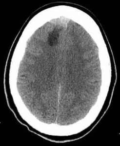
The arrow shows a low
density area in the frontal
lobe.
FEATURED BOOK: Neuroanatomy Through Clinical Cases (2nd ed.).
Because of the neutral density of the brain, it is easy to detect low or high density
changes. Injured brain tissue shows up on x-ray pictures as a low density change.
The term "low density" is typically used in CT (Computerized Tomography) scan reports.
CT scanning is an advanced imaging technique that uses x-rays and computer technology
to produces more clear and detailed pictures than a traditional x-ray. Low density is also
known as low attenuation. Density comes from the Latin word "densus" meaning "thick."
"Where Medical Information is Easy to Understand"™