MedFriendly®


Monocytes (High and Low Values)
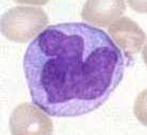
A monocyte (pictured below) is a large type of white
blood cell with one large, smooth, well-defined,
indented, slightly folded, oval, kidney-shaped, or
notched nucleus (the cells control center). White blood
cells help protect the body against diseases and fight
infections. The number of monocytes in the blood can
be detected with a test known as a complete blood
count (CBC) with differential. A CBC provides important
information about the kinds and numbers of cells in the
blood. Differential means that instead of only providing
the total white blood cell count that the different types
(aka differential) of white blood cells are listed.
A monocyte under a microscope.
FEATURED BOOK: Mosby's Diagnostic and Laboratory Test Reference
In the CBC test with differential, either the total number of monocytes is listed or the ratio
of the number of monocytes to the total number of white blood cells is listed. Knowing the
number of monocytes can help the health care provider rule in or rule out certain
diagnoses.
Monocytes are very flexible cells in that they can change depending on cues they receive
from the environment.
For example, they can develop into macrophages, which are cells that eat bacteria,
viruses, parasites, cells that have become infected, and debris in tissues.
"Where Medical Information is Easy to Understand"™
A parasite is an organism that lives in or on another organism to
obtain nourishment. Macrophages function at different locations
throughout the body once they are in the tissue. Macrophages
preserve an antigen so they can be recognized as foreign invaders
in the future. Antigens are substances in the body that can produce
a defensive reaction by the body. At times, macrophages function
as a scavenger type of cell and is why they are considered the big
eaters of the immune system. Macrophages are part of the innate
immune system. Monocytes (in macrophage form) serve as part of
what is known as the innate immune system of all mammals,
meaning that it immediately defends the body against infectious
agents in a general way.
In other words, they do not need to recognize specific types of invaders but generally recognize an
invader as something that must be destroyed.
Monocytes perform their functions by surrounding and engulfing bacteria (a process known as
phagocytosis). Monocytes can engage in phagocytosis by coating the foreign material with complement or
antibodies. Antibodies are types of proteins that are formed by the body to destroy foreign proteins known
as antigens (a process known as antibody-mediated cellular cytotoxicity). Complement is a type of protein
in the blood, which play a role in inflammation. Sometimes, monocytes attach to foreign materials by
recognizing them with specialized receptors. After phagocytosis, fragments of the foreign substance that
remains can serve as an antigen when the monocytes capture them and expose them to other white blood
cells known as T-cells, which leads to a specific response against it from the immune system. They
expose the fragments of the foreign substance with help from a special molecule known as an MHC (major
histocompatibility complex) molecule. Macrophages are also believed to play a role in forming important
organs such as the heart and the brain.
Monocytes can also divide into dendritic cells in the tissues. Dendritic cells are cells that process antigen
material and present it to the bodys immune (defense system). This is why they are considered a type of
antigen presenting cell. Unlike macrophages, dendritic cells do not destroy invaders directly but present
them to T cells (usually before they are fully developed) and B cells so they can learn more about them
and destroy them the next time they are encountered. T cells and B cells are types of small white blood
cells that help provide a specific response to attack the invading organisms and tumor cells. In dendritic
cell form, monocytes act as part of the acquired immune system (aka acquired immunity) in which highly
specialized cells protect the body against disease.
Monocytes respond to signals of inflammation in the body and can arrive quickly (about 8 to 12 hours) to
areas of infection or tissue damage and divide into macrophages and dendritic cells, which provides a
further immune system response. By responding to areas of tissue damage, they contribute to wound
healing. Macrophages also instruct other cells in the immune system and produce molecules that affect
the immune system.
Monocytes can also produce cytokines. Cytokines are proteins that help other white blood cells (and other
cells) communicate with each other. Names of cytokines usually produced by monocytes are interleukin-1,
interleukin-2,and tumor necrosis factor. When monocytes are activated, cytokines that promote
inflammation are produced and those which suppress inflammation are reduced. In the laboratory,
monocytes can be used to create dendritic cells by adding cytokines such as interleukin-4 and
Granulocyte Monocyte Colony Stimulating Factor (GMCSF).
Monocytes contain delicate chromatin material with a lacy or stringy pattern that appears more condensed
where the strands are in contact with each other. Chromatin is the material inside a nucleus from which
the chromosomes are formed. Chromosomes are structures in a person's cells that contain proteins and a
substance known as DNA (an abbreviation for deoxyribonucleic acid). DNA is a chain of many connected
genes. Genes are units of material contained in a person's cells that contain coded instructions as for
how certain bodily characteristics (such as eye color) will develop.
HOW BIG ARE MONOCYTES?
Monocytes are the largest type of blood cell. The size of monocyetes has been defined as between 13
and 25 micrometers in diameter, but some references state they are between 16 and 22 micrometers in
diameter. A micrometer is a very small unit of length that measures one millionth of a meter. A meter is
approximately 39 inches (slightly more than 3 feet).
WHERE ARE MONOCYTES MADE?
Monocytes are created by a type of cell in the bone marrow known as hematopoietic stem cells (HSCs),
which are cells that give rise to other cells. They create monoblasts which are premature forms of
monocytes. Monoblasts develop into monocytes.
WHAT PERCENT OF WHITE BLOOD CELLS ARE MONOCYTES?
Approximately 2-8% of white blood cells are monocytes. Monocytes move around in the blood for about
one to three days and the usually move into tissues throughout the body.
WHERE ARE MONOCYTES NORMALLY FOUND?
Monocytes are normally found in loose connective tissue, the spleen, lymph nodes, and bone marrow (a
tissue that fills the opening of bones). The spleen is an organ next to the stomach that helps fight infection
and removes and destroys worn-out red blood cells. Red blood cells are cells that help carry oxygen in the
blood. Incidentally, some monocytes phagocytize red blood cells. Lymph nodes are small egg shaped
structures in the body that help fight against infection. About half of the monocytes are found in the spleen,
in an area of connective tissue known as the cords of Billroth.
Macrophages are found in various forms in all tissues of the human body. These forms include microglia,
osteoclasts, histiocytes, and Kupffer cells. Microglia are macrophages in the brain and spinal cord.
Osteoclasts are types of bone cells that remove bone tissue. A histiocyte is a type of macrophage of the
immune system. Kuppfer cells are types of macrophages in the liver. The liver is the largest organ in the
body and is responsible for filtering (removing) harmful chemical substances, producing important
chemicals for the body, and other important functions.
HOW DO MONOCYTES RESPOND TO LABORATORY STAINING?
When stained with dyes, monocytes display a significant amount of pale blue or blue-gray cytoplasm.
Cytoplasm is all the substance of a cell except the nucleus and cell wall. Monocvtes have a large amount
of cytoplasm. The cytoplasm of monocytes is filled with many fine, dust-like, reddish-blue granules.
Vacuoles are often present, which are clear spaces in the substance of a cell. This is typically the case
when the monocyte has phagocytized a foreign substance. Monocytes also contain many vesicles (small
bubbles within a cell) for processing foreign materials. A picture of a monocyte is shown below.
WHAT ARE THE DIFFERENT TYPES OF MONOCYTES?
There are at least three different types of monocytes located in the blood of humans. The first is known as
a classical monocyte, which has a high amount of CD14 receptors on the surface. The CD14 receptor
helps detect certain types of bacteria molecules. There is a non-classical monocyte, which has a low
amount of CD14 receptors on the surface but has some CD16 receptors, which contributes to the
protective function of the immune system. There is also an intermediate monocytes which has a high level
of CD14 receptors and a low level of CD16 receptors.
The classical monocyte (which does not have CD 16 receptors) appears to develop into intermediate
monocytes and then into the non-classical monocyte. This is why the non-classical monocyte is
sometimes viewed as more mature type of monocyte. When stimulated, the intermediate monocytes
produce high amounts of cytokines that promote inflammation (e.g., interleukin-12 and tumor necrosis
factor). A protein known as PD-1 (programmed cell death 1) is found more in intermediate monocytes than
classical monocytes. PD-1 interacts with another molecule (known as PD-L1) to prevent overreaction of
the immune system.
WHAT CAUSES THE MONOCYTE LEVEL TO BE TOO HIGH?
When the monocyte level is too high, this is known as monocytosis. This can happen for several reasons
such as stress, inflammation, a fever from a virus, severe infection (because more macrophages are
needed to fight it), premature cell death in living tissue, diseases that result from abnormal activity of the
immune system, and regeneration of red blood cells.
Another cause of high monocytes is sarcoidosis. Sarcoidosis is a condition in which small, rounded
bumps form on tissues. Chronic granulomatous disease (CGD) can also cause monocytosis. CGD is an
inherited disorder in which cells that perform phagocytosis do not function correctly. Another cause of high
monocytes is Cushings syndrome, a condition caused by excessive exposure to high levels of cortisol.
Cortisol is a type of hormone produced by the adrenal glands that helps reduce inflammation. Hormones
are types of chemicals in the body that affect other cells. The adrenal glands are a pair of glands that play
an important role in metabolism and help the body respond to physical and emotional stress by releasing
certain hormones. Metabolism is the chemical actions in cells that release energy from nutrients or use
energy to create other substances.
WHAT CAUSES THE MONOCYTE LEVEL TO BE TOO LOW?
When the monocyte level is too low, this is known as monocytopenia. Because monocytes are produced
in the bone marrow, any illness or chemical that affects the bone marrow can cause a low monocyte
count. There are many health conditions that affect the bone marrow. One example is HIV (human
immunodeficiency virus). HIV is a virus that attacks the bodys immune system, leading to infections and
harmful tumors. Tumors are abnormal masses of tissue that form when cells in a certain area of the body
reproduce at an increased rate.
Another example of a condition that can affect bone marrow is aplastic anemia. Anaplastic anemia is a
condition in which the body stops producing enough blood cells due to bone marrow damage. Yet another
example is systemic lupus erythematosus (SLE). SLE is a long-term disease in which the connective
tissues throughout the body are inflamed because the body's defense system attacks these tissues as if
they were foreign substances.
Another example is rheumatoid arthritis. Rheumatoid arthritis is an immune disorder in which the body's
defense system attacks its own tissues, causing inflammation of bone joints. Other examples include
tuberculosis, malaria, and Epstein-Barr virus. Tuberculosis is a highly infectious diseases characterized
by the formation of small swellings called nodules (or tubercles) in tissues, especially the lungs. Malaria is
a serious disease caused by parasites that is spread by mosquitoes. The Epstein-Barr virus is one of the
most common viruses in humans.
Medications that can interfere with bone marrow and lower the monocyte count includes chemotherapy,
radiation therapy, interferons, and glucocorticoids This can be caused by therapy with glucocorticoids,
which are types of steroid hormones that suppress the immune system. Chemotherapy is a type of
medication used to treat cancer. Cancer is an abnormal growth of new tissue characterized by
uncontrolled growth of abnormally structured cells that have a more primitive form. Radiation is a type of
energy that is sometimes aimed at cancer tumors to destroy or weaken them. Interferons are proteins
released in the body in response to the presence of harmful substances (such as tumor cells) which then
trigger the immune system to fight them. Low monocyte counts can also be caused low levels of folic acid
(a type of vitamin) and vitamin B12.
WHAT IS THE ORIGIN OF THE TERM "MONOCYTE"?
Monocyte comes from the Greek word "monos" meaning "single," and the Greek word "kytos" meaning
"cell." Put the two words together and you have "single cell."