


MedFriendly®


Periventricular
White Matter
White Matter
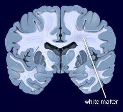
Periventricular white matter refers to white
matter that is immediately to the side of the
two lateral (side) ventricles of the brain. This
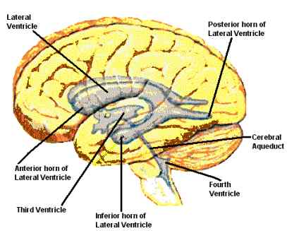
is shown in the second picture below. The
lateral ventricles are two curved openings
(shaped like a horseshoe) located deep
within the top section of the brain.
White matter is a group of white nerve fibers
that conduct nerve impulses quickly. White
matter is important for muscle movements.
FEATURED BOOK: Neuroanatomy Through Clinical Cases (2nd ed.)
ARE THERE DIFFERENT AREAS OF PERIVENTRICULAR WHITE MATTER?
Yes. Periventrivcular white matter is categorized based on which lobe (section) of the
brain that it is located. There are the four main lobes of the brain, the frontal lobes,
occipital lobes, temporal lobes, and parietal lobes.
The frontal lobes are located in the front of the brain and the occipital lobes are located
in the back of the brain. The temporal lobes are located on the sides of the brain behind
the ears. The parietal lobe is the middle area of the top part of the brain. Periventricular
white matter located near the frontal lobes is called frontal periventricular white matter.
Periventricular white matter located near the occipital lobes is called occipital
periventricular white matter.
"Where Medical Information is Easy to Understand"™
Periventricular white matter located near the temporal lobes is called
temporal periventricular white matter. Periventricular white matter
located near the parietal lobes is called parietal periventricular white
matter.
WHERE ARE THE VENTRICLES LOCATED?
As can be seen in the picture above, the ventricles are locate
throughout the brain and they are all connected. That is a view from
the side of the brain looking inwards.
WHAT ARE PERIVENTRICULAR WHITE MATTER CHANGES?
Periventricular white matter changes means that there has been some change in the structure of the
white matter near the ventricles of the brain. This finding does not necessarily mean that something
serious, like a disease, has caused it. In fact, the most common cause of periventricular white matter
changes is normal aging that is not associated with a disease process.
In premature infants (babies born too early), however, the periventricular white matter is a common area
of damage when an event happens that causes a lack of oxygen to the brain. Periventricular white matter
changes in premature infants typically occurs near the collateral trigone. The collateral trigone is a
triangle-shaped prominence on the floor of the lateral ventricle.
In people over age 65, research has shown that periventricular white matter changes are found between
30% and 80% of the time when an MRI scan of the brain is performed. MRI stands for Magnetic
Resonance Imaging. MRI scans produce extremely detailed pictures of the inside of the body by using
very powerful magnets and computer technology.
Possible causes of periventricular white matter changes include Binswanger's disease, stroke, migraine
headaches, multiple sclerosis, and CADASIL (Cerebral Autosomal Dominant Arteriopathy with Subcortical
Infarcts and Leukoencephalopathy). Each of these terms are described below.
Binswanger's disease is a type of dementia in which white matter below the
cortex (the top, main section of the brain) wears away and becomes thinner.
Dementia is a mental disorder characterized by a significant loss of
intellectual and cognitive abilities without impairment of perception or
consciousness. A stroke is a burst artery (a type of blood vessel that carries
blood away from the heart) or a blockage of an artery in the brain. A migraine
headache is a painful and continuous type of headache that is so painful that
it prevents people who experience them from doing anything. Multiple
sclerosis is a condition in which multiple areas of abnormal patches (known
as plaques) develop in the brain and/or spinal cord. ICADASIL is a rare,
inherited disease starting in mid-adulthood that causes abnormal changes to
many small arteries in the brain.
People with periventricular white matter changes usually perform in the low average range or worse on
tests of psychomotor speed. In general, however, periventricular white matter changes are not
associated with any particular set of symptoms. Thus, one cannot be sure that a patient's difficulty
walking or other problems are caused by periventricular white matter changes.
WHAT IS THE ORIGIN OF THE TERM, PERIVENTRICULAR WHITE MATTER?
Periventricular white matter comes from the Greek word "peri" meaning "around," the Latin word "venter"
meaning "belly," the Proto-Indo-European word "kwintos" meaning "bright," and the Latin word "materies"
meaning "substance." Put the words together and you have "bright substance around (the) belly."
