MedFriendly®


Villus
In general, a villus is a tiny, thin, fingerlike structure with
a blood supply that sticks out from the surface. More
than one villus is known as villi. Villi are located in
different areas of the body. Most commonly, the term is
used to describe the many tiny, fingerlike structures
that stick out and are located in groups over the entire
mucous surface (a type of thin sheet of tissue) of the
small intestine. The intestine is a tube shaped structure
that is part of the digestive tract. The small intestine is
a part of the intestine that takes in all of the nutrients
(healthy substances) that the body needs.
FEATURED BOOK: Breaking the Vicious Cycle: Intestinal Health through Diet
The villi help to increase the total area on the outside of the small intestine to the size of
about half a tennis court. In this way, the villi help absorb, move, and distribute some of
the fluids and nutrients into the blood and lymphatic system. The lymphatic system is a
system of vessels that drain lymph from all over the body back into the blood. Lymph is a
milky fluid that contains proteins, fats, and white blood cells (which help the body fight off
diseases). Food particles that are broken down in the digestive system reach the blood
through the capillaries (very tiny blood vessels) in the villi.
CAN A VILLUS BE SEEN?
Yes, although a villus can barely be seen with the naked eye.
"Where Medical Information is Easy to Understand"™
HOW MANY VILLI ARE IN THE BODY?
There are millions of villi in the body. In fact, there are millions of villi
on the small intestine.
These villi are the largest and most present on the first and second
parts of the small intestine, which are known as the duodenum and
jejunem.
These two areas are where most of the absorption of food occurs.
The villi become more flat when the intestine widens.
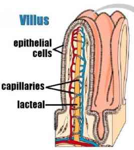
WHAT IS A VILLUS MADE OF?
As you can see in the picture above, a villus is covered by types of cells known as epithelium. Cells are
the smallest, most basic units of life, that are capable of existing by themselves. It is the epithelial cells
that actually help absorb, move, and distribute some of the fluids and nutrients in the body.
You can also see that the villus has a network of capillaries, which are very tiny blood vessels. The villus
also has another type of tiny vessel known as a lacteal, which fills with milky white fluid known as chyle
after a fatty meal. Each villus is supported by types of delicate tissue known as areolar and reticular
connective tissue. These tissues support the epithelial cells, mentioned above.
WHAT ARE SOME OTHER PLACES THAT VILLI ARE LOCATED?
Before they are born, when babies are in the earliest stages of development, villi are found on the surface
of the outermost membrane (a thin layer of tissue) of the organism. This outermost layer is known as the
chorion. These type of villi are known as chorionic villi. Chorionic villi help form the placenta, an organ in a
female that nourishes a baby during pregnancy. Villi are also located in the brain. In this case, the villi are
types of tissue that stick out from one of the layers that cover the brain. This layer is known as the
arachnoid membrane, which is why these villi are known as arachnoid villi.
WHAT IS VILLOUS BLUNTING?
Villous blunting is when the villu have become dull or less defined.
DOES THE TERM "VILLUS" HAVE ANY OTHER MEANINGS?
Yes. Villus is also used to mean a long bump in the skin that goes into an abnormal opening or a fluid-filled,
blister-like opening within the top layer of the skin.
WHAT IS THE ORIGIN OF THE TERM, VILLUS?
Villus is the Latin word for "shaggy hair."