


MedFriendly®


Angiography
An angiography is a technique that produces a
picture of the inside structure of blood vessels and/or
the heart by using radiation (a type of energy) known
as X-rays or gamma rays. The blood vessels typically
examined with an angiography are arteries and veins.
An artery carries blood away from the heart, whereas
a vein carries blood to the heart. Angiographic means
pertaining to or using an angiography.
HOW DOES RADIATION PRODUCE A PICTURE?
After radiation hits the body, it is absorbed by
different body parts in different ways. The body part
that is the focus of the radiation casts a shadow on
film.
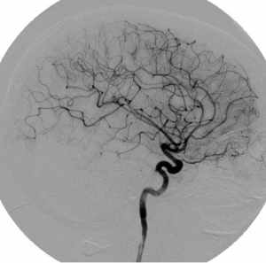
Blood vessels pictured during
an angiography.
This shadow leads to a picture of the body part, with the bones appearing white and the
soft tissues appearing dark gray. In an angiography, the blood vessels are filled with a
substance (known as a contrast) that radiation cannot pass through.
This allows the blood vessels to be seen in contrast to the surrounding areas that the x-
rays can pass through.
FEATURED BOOK: Abrams' Angiography: Interventional Radiology
"Where Medical Information is Easy to Understand"™
WHY IS AN ANGIOGRAPHY PERFORMED?
Angiography is performed to detect diseases that change the
appearance of the inside of blood vessels. Some diseases can
cause the formation of an aneurysm. An aneurysm is a balloon-like
expansion of a blood vessel due to weakening of the blood vessel
walls. The expanded blood vessel can burst if it becomes large
enough.
Other diseases involve a narrowing or blockage of the blood vessel
walls. One cause of such narrowing or blockage includes a buildup
of patchy areas known as calcified artherosclerotic plaques.
Angiograms are also used to assess changes in blood vessels caused by tumors (tissues that grow more
rapidly than normal) and damage to organs. By assessing the changes in the blood vessels, the doctor
can determine the extent of disease and plan treatment based on the results of the assessment.
Other conditions that angiographies are used to diagnose are heart attacks, stroke (a burst artery or a
blockage of an artery in the brain), kidney tumors, narrowing of the renal artery (a pair of arteries in the
kidneys), and increased blood pressure in the portal vein. The portal vein is a large vein that carries blood
from the stomach, intestine, and spleen to the liver. The intestine is a tube shaped structure that is part of
the digestive tract. The spleen is an organ that helps fight infection and removes and destroys worn-out
red blood cells.
HOW IS AN ANGIOGRAPHY PERFORMED?
First, the skin and tissues around a blood vessel (artery or vein) are numbed with a medication so the
patient does not feel the needle being inserted into the skin. Next, a long thin wire with a soft tip is
inserted through the needle and into the blood vessel. This wire is known as a guide wire. With the guide
wire in place, the needle is removed. A flexible plastic tube (known as a catheter) is then placed over the
guide wire. The guide wire helps move the tip of the catheter into the desired location of the blood vessel
that needs to be examined. The doctors can tell where the catheter is by using pictures produced by x-
rays.
Once the catheter reaches the desired blood vessel, the contrast material (described in the 2nd question)
is injected into the blood vessel. A rapid sequence or movie of x-ray pictures is studied to monitor the flow
of contrast through the blood vessels. In this way, the blood vessels are examined. The contrast material
is usually inserted in the femoral artery (a large artery in each leg), the carotid artery (located on both
sides of the neck), or the brachial artery (located over each elbow).
HOW LONG DOES AN ANGIOGRAPHY TAKE?
An angiography can last anywhere from a few minutes to as long as two to three hours.
ARE THERE DIFFERENT TYPES OF ANGIOGRAPHIES?
Yes. There are many different types of angiographies, each of which examine blood vessels in different
areas of the body. Perhaps the most well known angiography is a coronary angiography, which is done to
assess if there is any narrowing or blockage of arteries near the heart. A carotid angiography is
sometimes used to examine if there is a narrowing or blockage in one of the carotid arteries. The carotid
arteries are located in the neck and supplies blood to the brain. A cerebral angiography is used to
determine if an aneurysm (see last question for description) is present in the brain. It is also used to
visualize a brain tumor before surgery. A special type of angiography that does not use a catheter and
does not require the use of contrast is known as a magnetic resonance angiography.
WHAT RISKS ARE INVOLVED WITH AN ANGIOGRAPHY?
The contrast material (see above) that is injected in the blood vessel usually causes a sensation of
warmth throughout the body. However, this sensation typically lasts only for a few seconds. The warm
sensation may be greater in the part of the body where the injection took place. A more serious side
effect occurs when someone is allergic to an ingredient in the contrast material. Such ingredients include
iodine (a nonmetallic element) and shellfish. Thus, it is recommended that patients be tested for allergic
reactions to the contrast before the procedure is performed. However, it should be noted that with
improvements in the development of contrasts, less than one person in 80,000 will be allergic to the
contrast used in an angiography. Another risk is that blood vessels can potentially be damaged during an
angiography since a needle, a wire, and a catheter (flexible, plastic tube) is being placed in them. This is
why the patient is recommended to rest in bed after the procedure and be monitored for signs of bleeding.
ARE ANGIOGRAPHIES ALSO USED FOR TREATMENT?
Yes. Angiographies are also performed to assist in certain types of treatment that can eliminate the need
for surgery. For example, in cases where the inside of the artery is narrowed, a small balloon can be
inflated at the tip of the catheter to widen this area. This procedure is known as a balloon angioplasty.
Another form of treatment that angiographies are used for is to inject material that reduces or shuts off
the blood supply to tumors (tissues that grow more rapidly than normal). By using an angiography,
medication can also be sent directly to blood vessels to control bleeding. Angiographies can also be used
to send medication directly into the blood supply to individual organs.
WHAT IS THE ORIGIN OF THE TERM, ANGIOGRAPHY?
Angiography comes from the Greek word "angeion" meaning "a vessel or opening in the body," and the
Greek word "graphe" meaning "to write." Put the two words together and you get "to write (about) a vessel
or opening of the body.