MedFriendly®


Ganglioglioma
A ganglioglioma is a rare type of brain tumor. Tumors are
issues that grow more rapidly than normal. Gangliogliomas are
types of brain tumors that are made of mixed groups of cells.
A cell is the smallest, most basic unit of life, that is capable of
existing by itself. Gangliogliomas are partly made of abnormal
glial cells. Glial cells are cells that support and maintain other
cells. Gangliogliomas are also partly made of neurons (nerve
cells) that are in various degrees of abnormality. Whereas
neurons transmit and receive information to and from one
another, glial cells do not.
Supportive tissue known as stroma, which contains fibers and
blood vessels, are also found in gangliogliomas. Fibers are
flexible, threadlike objects found outside of cells.
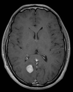
A ganglioma on a brain MRI.
FEATURED BOOK: I Have a Life to Live! Coping with a Brain Tumor
Blood vessels are tube shaped structures that carry blood to and from the body. In
gangliogliomas, the neurons, glial cells, and stroma are abnormal in shape, size, and
appearance.
Rarely, star-shaped types of glial cells known as astrocytes can dramatically change in
structure, going back to a very early form of development. As a general rule, the more the
cells change in structure, size, and appearance, the more harmful the tumor is.
"Where Medical Information is Easy to Understand"™
WHAT ARE SOME OTHER CHARACTERISTICS OF
GANGLIOGLIOMAS?
Gangliogliomas are gray in appearance and feel firm when removed
from the brain. About 50% of gangliogliomas are associated with
cysts. A cyst is an abnormal lump, swelling, or sac that contains
fluid, a part solid material, or a gas, and is covered with a
membrane. A membrane is a thin layer of flexible tissue that covers
something. Cysts are present in and under the skin. About 35 to
50% of all gangliogliomas have a buildup of calcium associated with
them. Calcium is a natural element that is very important in bone
formation.
Five percent of patients with gangliogliomas have additional abnormalities such as a total absence of the
corpus callosum. The corpus callosum is a large band of nerve fibers in the brain that help the two sides of
the brain communicate with each other. Another type of abnormality that is rarely associated with
gangliogliomas is Down syndrome. Down syndrome is an abnormality present from birth that results in
mental impairments and a characteristic physical appearance (small facial features, large tongue that
sticks out, a flat back area of the head, and hands that are short and broad).
WHERE DO MOST GANGLIOGLIOMAS OCCUR?
Gangliogliomas can occur anywhere in the brain and rarely occur in the spine. Most gangliogliomas are
located in the temporal lobes. The temporal lobes are located on the sides of the brain by the ears. 70%
of gangliogliomas in children are located above a part in the brain known as the tentorium. The tentorium
is a membrane that separates an area in the back, lower part of the brain known as the cerebellum, from
an area in the back, upper part of the brain known as the occipital lobes. Five to ten percent of child brain
tumors that are found above the tentorium are gangliogliomas. Gangliogliomas can also occur in the
brainstem, which is an area located below the cerebellum that controls many important motor, sensory,
and reflex actions.
It is worth noting, however, that are still a significant number of gangliogliomas that are present below the
tentorium, in the cerebellum. The cerebellum plays an important role in movement and coordination. More
unusual locations for gangliogliomas to be found are in the basal ganglia, hypothalamus, optic chiasm, and
pineal gland. The basal ganglia are paired groups of nerve cells located deep within the brain that play an
important part in smooth, continuous muscle movements and in starting and stopping movements. The
hypothalamus is an area located in the lower part of the brain that is important for many bodily functions
such as sleep, thirst, and hunger. The optic chiasm is an area in the brain where optic nerves (nerves that
help us see) cross over. The pineal gland is a pine cone-shaped structure in the lower part of the brain
that produces melatonin. Melatonin is a natural chemical in the body thought to promote sleep.
WHAT ARE THE SIGNS AND SYMPTOMS OF GANGLIOGLIOMA?
The signs and symptoms of gangliogliomas depend on the location of the tumor, how fast it spreads, and
the age of the patient. In people ages 10 to 20, gangliogliomas are often associated with seizures, which
are involuntary muscle movements and/or decreased awareness of the environment due to
overexcitement of nerve cells in the brain. Since most gangliogliomas appear in the temporal lobes, many
people with this type of tumor develop seizures that come from the temporal lobes. Seizures coming from
the temporal lobes have many different features, but usually include a loss of awareness (but not always),
abnormal sensations, feeling detached from oneself, and involuntary muscle movements.
If the ganglioglioma is present is the cerebellum, different signs and symptoms will be present such as
impaired motor coordination, headache, and a buildup of cerebrospinal fluid in the brain. Cerebrospinal
fluid bathes and cushions the outside of brain and spinal cord. If the hypothalamus is affected, problems
associated with functions that this area of the brain plays an important role in (for example, sleep, thirst,
and hunger) can be present. These problems are usually found in people ages 10-20 that have
gangliogliomas.
HOW ARE GANGLIOGLIOMAS DIAGNOSED?
Many patients with gangliogliomas go to the doctor because of the seizures that are usually associated
with them. This leads the doctor to use various techniques to diagnose the cause of the seizures.
Gangliogliomas are partly diagnosed with techniques that provide pictures of the brain. One such
technique is a CT (computerized tomography) scan. CT scanning is an advanced imaging technique that
uses x-rays and computer technology to produces more clear and detailed pictures than a traditional x-
ray.
Magnetic resonance imaging (MRI) scans of the brain are often used. MRI scans produce extremely
detailed pictures of the inside of the body by using very powerful magnets and computer technology. MRI
scans are more detailed and more expensive that CT scans. MRI scans are the most sensitive and
specific at detecting that a tumor is present, particularly when contrast is injected into the person's body
during the scan. Contrast is a substance that allows damaged areas to show up better on the picture
produced by the MRI. About 50% of gangliogliomas will be detected when contrast is used.
MRI scans are also the best at detecting if there are any cysts or other solid materials associated with
the gangliogliomas. A cyst is an abnormal lump, swelling, or sac that contains fluid, a part solid material, or
a gas, and is covered with a membrane. A membrane is a thin layer of flexible tissue that covers
something. Cysts are present in and under the skin.
Unfortunately, gangliogliomas do not have a very specific appearance on CT or MRI scans. Thus, when a
ganglioglioma is present, all that doctors usually know before surgery is that there is a tumor in the brain,
but not what type. Nonetheless, if a person has a tumor in the temporal lobes without edema (a buildup of
watery fluid) surrounding it, and seizures that will not respond to medications, it is likely that the tumor is a
ganglioglioma. It is not until a sample of the tumor is removed during surgery that it can be tested in a
laboratory and definitively stated that it is a ganglioglioma. This diagnosis is based on certain
characteristic features of the tumor, some of which are listed below.
In the laboratory, it is found that the abnormal neurons are extremely reactive to anything that may irritate
them. Only after the neurons are stimulated from irritation will a protein known as synaptophysin can be
found in them. Another type of protein that is found in gangliogliomas is called GFAP (glial fibrillary acidic
protein). In order to detect the presence of these proteins, special stains needs to be applied to the
neurons in the lab. Another characteristic of abnormal neurons in gangliogliomas is that they have two
nuclei instead of one. A nucleus is a structure in the center of a cell. You can see a picture of a nucleus in
our entry for cell. More than one nucleus are called nuclei.
DO REGULAR X-RAYS HELP DIAGNOSIS GANGLIOGLIOMAS?
Regular x-rays usually do not help in diagnosing gangliogliomas because they are not good at detecting
tissue. X-rays are best at detecting bones. However, sometimes enough calcium may form near the
ganglioglioma to be detected on x-rays. Calcium is a natural element that is important is bone formation.
WHAT IS THE AGE OF MOST PEOPLE WHEN DIAGNOSED WITH GANGLIOGLIOMA?
Gangliogliomas are most common before age 20. About 60% of people who get diagnosed with
gangliogliomas are adolescents and adults younger than age 30. However, people as young as 2 and as
old as 70 have been known to develop gangliogliomas.
DO MORE MALES OR FEMALES DEVELOP GANGLIOGLIOMAS?
Approximately equal numbers of males and females develop gangliogliomas.
DOES ONE PARTICULAR RACE DEVELOP GANGLIOGLIOMS MORE THAN ANOTHER?
No. No particular race has been shown to be more likely to develop gangliogliomas.
HOW FREQUENT ARE GANGLIOGLIOMAS?
In the United States, approximately 1 to 2% (with a range of about 0.4 to 7.6%) of brain tumors are
gangliogliomas. One percent of tumors inside the spinal cord are gangliogliomas. About 10% of all primary
brain tumors in children are gangliogliomas. Primary brain tumors are tumors that originated in the brain as
opposed to spreading to the brain from somewhere else in the body, such as the chest.
HOW ARE GANGLIOGLIOMAS TREATED?
Gangliogliomas are usually treated with surgery to remove it. After the surgery is performed, radiation
therapy is not needed unless the tumor has shown signs of growing back, which would be rare. Radiation
is a type of energy that is sometimes aimed at tumors to destroy or weaken them.
WHAT IS THE PROGNOSIS OF PEOPLE WITH GANGLIOGLIOMAS?
The prognosis of people with gangliogliomas is excellent if it has been totally removed. The excellent
prognosis in people with gangliogliomas is helped by the fact that most of these tumors are not
aggressive, meaning that they do not spread very fast. Thus, it is usually easy for surgeons to remove
these tumors because they are typically found in one place and can be separated from the brain tissue.
Most people are cured after such surgery and the chances of the tumor coming back after it has been
totally removed is rare. The ten-year survival rate in children who have been treated with ganglioglioma is
about 90%. Prognosis of people with gangliogliomas is better the earlier it is diagnosed and treated.
WHO FIRST DESCRIBED GANGLIOGLIOMAS?
Gangliogliomas were first described by CB Courville in his 1930 paper, "Ganglioglioma; tumor of central
nervous system. Review of the literature and report of 2 cases," published in the Archives of Neurology
and Psychiatry, Volume 24, pages 439-491. He described gangliogliomas as abnormal growths of new
tissues that contained neurons (nerve cells) and types of glial cells known as astrocytes. See the
beginning of this entry for more on the difference between neurons and glial cells.
WHAT IS THE ORIGIN OF THE TERM, GANGLIOGLIOMA?
Ganglioglioma comes from the Greek word "ganglion" meaning "knot," the Greek word "glia" meaning
"glue," and the Greek word "oma" meaning "tumor." Put the words together and you get "glue knot tumor."