MedFriendly®


Hypoxic Ischemic Encephalopathy
HIE) means permanent brain injury caused by a lack of
oxygen or below normal levels of blood flow to the
brain. The brain needs a constant supply of oxygen in
order to function properly. If oxygen fails to flow from
the arteries (blood vessels that carry blood away from
the heart) to the brain tissue, or if there is a decreased
amount of oxygen in the blood of the arteries, HIE can
result. HIE is one of the most common and severe
types of brain injury seen in emergency rooms and
recovery rooms of virtually every general hospital.
WHAT HAPPENS TO THE BRAIN WHEN OXYGEN
BEGINS TO BE REDUCED?
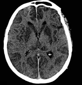
This brain CT scan shows signs of
hypoxic ischemic encephalopathy.
FEATURED BOOK: Neuroanatomy through Clinical Cases
When the oxygen supply to the brain is reduced, the body tries to get more blood to it by
widening blood vessels, so more blood can flow through at a constant rate. However, this
defense strategy of the body does not work for long (see below). If the person is awake,
he/she will likely have poor attention, poor judgment, and poor coordination of muscles.
WHAT CAUSES HYPOXIC ISCHEMIC ENCEPHALOPATHY?
Hypoxic ischemic encephalopathy is most often caused by the following:
"Where Medical Information is Easy to Understand"™
HEART ATTACK
IRREGULAR HEARTBEAT
SHOCK FROM A TRAUMA
MASSIVE BLEEDING: Either on the outer surface of the body or in
the organs inside of the body.
SUFFOCATION: From drowning, choking, being strangled, or the
presence of something in the windpipe (such as blood or a foreign
object) that prevents breathing.
CARBON MONOXIDE POISONING: Poisoning by a gas called carbon monoxide. Carbon monoxide
prevents the blood from receiving enough oxygen. Breathing stops first, then the heart stops.
PROBLEMS WITH HEMOGLOBIN: Hemoglobin is a substance that carries oxygen in the blood. If this
substance is not functioning properly, there will not be enough oxygen in the blood that reaches the brain.
SEPTIC SHOCK: A form of shock when poisons are released from harmful organisms (known as bacteria)
in the blood. The bacteria are in the blood because they have spread from an infection somewhere else in
the body.
GENERAL ANESTHESIA: The use of substance to prevent sensation. This is generally done before
many types of surgery so that the patient does not feel pain. While being operated on, the patient
receives air to breathe. If the air does not have enough oxygen, HIE can result.
DISEASES THAT PREVENT THE MOVEMENT OF MUSCLES THAT HELP IN BREATHING: The names
of diseases that can do this are amyotrophic lateral sclerosis (ALS), myasthenia, and Guillain-Barre
syndrome. A disease called poliomyelitis (also known as polio) can also cause this to happen, but the
disease is not seen much today.
BRAIN DAMAGE: Damage to an area in the lower part of the brain that controls breathing, known as the
medulla. This damage can be caused by a trauma such as a car accident, bleeding or a lack of oxygen in
the area of the medulla, and epilepsy. Epilepsy is a brain condition that is characterized by changes in
sensation, abnormal behaviors, loss of consciousness, convulsive seizures, or all of these. Convulsive
seizures are sudden abnormal, severe involuntary muscle movements, with a racing heart and other
symptoms.
WHAT HAPPENS NEXT & HOW LONG DOES IT TAKE BEFORE THE TISSUE DIES?
If the oxygen supply continues to be reduced, the cells in the brain will break down in an attempt to
produce energy. In the process of breaking down, the cells cause such damage to themselves, that they
die. Massive amounts of calcium (a metallic element) present in the body are believed to flow into the
tissue area in large amounts and assist in the death of the cells. It takes about five minutes (sometimes
longer), for a tissue to die that is not receiving enough oxygen. Tissues are made up of groups of cells.
HOW IS HYPOXIC ISCHEMIC ENCEPHALOPATHY TREATED?
The purpose of treatment for HIE is to prevent serious injury from occurring. To do this, the flow of
oxygen, as well as proper heart and lung functioning needs to be restored immediately. To restore heart
functioning, an electric shock may need to be applied to the heart. If breathing, oxygen flow in the blood,
and heart functioning is restored within 3 to 5 minutes, then recovery can occur. However, if the oxygen
blood flow is cut off for more than 5 minutes, permanent brain injury usually results.
Increased blood pressure, heart action, and low body temperatures may help the person resist permanent
injury if the blood flow is cut off for slightly more than 5 minutes. However, breathing, oxygen flow in the
blood, and heart functioning must be restored for recovery to occur.
WHAT IS COMMONLY SEEN IN PATIENTS WHO RECOVER FROM A BRIEF DECREASE IN OXYGEN
TO THE BRAIN?
As was mentioned earlier, recovery can occur from a decrease in oxygen to the brain, if the oxygen
supply is restored within three to five minutes. Typically, such a patient is in a coma for a few days. A
coma is a state of deep unconsciousness in which there are no voluntary movements, no responses to
pain, and no verbal speech. Eventually, consciousness is regained, which is followed by confusion,
abnormal movements, and an inability in recognizing visual objects even though vision is intact. Some
patients pass through this phase quickly and make a full recovery. However, others are left with different
degrees of permanent disability from brain damage (see below).
WHAT ARE SOME OF THE PERMAMENT DISABILITIES THAT CAN OCCUR FROM BRAIN DAMAGE
DUE TO A LACK OF OXYGEN?
Permanent disabilities that can occur from brain damage due to a lack of oxygen include the following.
Please note that these disabilities often overlap in various combinations, although any one of them may
be the main disability experienced.
• Abnormal movements.
• Long-term coma (see last section).
• Seizures: sudden, violent, involuntary muscle movements.
• Inability in recognizing visual objects even though vision is intact.
• Loss of muscle coordination due to damage to an area in the back of the brain known as the cerebellum.
• Rapid, uncontrollable jerking muscle movements that occur at rest or at movement.
• Choreoathetosis: abnormal body movements characterized by a choreic (involuntary, irregular, dance-
like, movements of the arms, legs, and face) and athetoid (involuntary, slow, twisting movements of the
hands, fingers, toes, and feet) pattern.
• Dementia: a mental disorder that gets worse over time and is characterized by confusion, impaired
thinking abilities, poor memory, poor judgment, personality changes, unawareness of the environment,
unresponsiveness, and doing things based on impulse.
• Disorientation (confusion), severely impaired memory of recent events (also known as anterograde
amnesia, and an inability to learn new skills.
There are some patients that will make a recovery and then get worse after a period of 1 to 4 weeks.
These patients usually feel confused, irritable, and experience a lack of interest in things. Some patients
feel agitated and high-strung. This worsening of symptoms is uncommon and not well understood. It is
known as delayed postanoxic encephalopathy. Many patients survive it, but some are left with serious
impairments in thinking and movement. Some patients continue to worsen and die within 1 to 2 weeks.
After death, such patients are found to have experienced widespread destruction of myelin in the brain.
Myelin is a substance that makes up the tube-shaped covering of various nerve fibers in the body and
helps them transmit messages fast.
There are also some patients that slowly get worse over weeks and months until they stop speaking, are
stiff, and cannot help themselves in any way. In such patients, the damage is mostly located in an area
known as the basal ganglia. The basal ganglia are paired groups of nerve cells located deep within the
brain that play an important part in smooth, continuous muscle movements and in starting and stopping
movements.
WHAT ARE SIGNS THAT CAN HELP TELL IF SOMEONE WILL RECOVER FROM HYPOXIC ISCHEMIC
ENCEPHALOPATHY?
The doctor or nurse should look for signs that the brainstem is intact. The brainstem is an area in the
lower part of the brain that connects it with the spinal cord. This area of the brain controls many functions
crucial for life to continue. One such sign includes the pupil of the eye widening in response to light. The
pupil is the dark circle in the middle of the eye. Another sign is the ciliospinal reflex, in which the pupils
widen by scratching or pinching the skin of the neck or face.
Yet another sign is called "doll's head eye movements": If the brain stem is functioning normally, the eyes
will move in the opposite direction when quickly rotated from side to side. If the signs mentioned above
are not present, especially the widening of the pupils in response to light, the patient's condition is likely
hopeless.
The most severe cases of HIE are those in which the responses described above are not present, the
patient is totally unaware of his/her environment, natural breathing stops, and there is no electrical activity
in the brain detected with tests. If severe, continuing seizures (sudden, violent, involuntary muscle
movements) occur that do not respond to treatment, a poor outcome is likely.
DO ANY PATIENTS SURVIVE SEVERE HYPOXIC ISCHEMIC ENCEPHALOPATHY?
Yes. However, some patients do not speak afterwards, will not respond to others, and are unaware of
their environment for weeks, months, or years. Some improvement may occur over the long-term, but the
patient appears not to know anything about the present, has lost the ability to reason, live independently,
recall memories from the past, and interact with others in a socially acceptable way.
WHAT HAPPENS TO PATIENTS WHO HAVE BRAIN DEATH FROM SEVERE HYPOXIC ISCHEMIC
ENCEPHALOPATHY?
In patients with severe HIE, in which brain death occurs, the decision must be made as to whether to end
life support. However, doctors must be very careful in stating that brain damage cannot be reversed
because anesthesia (see above), drug overdose, and severely low body temperature can appear like
irreversible brain damage. When properly treated however, recovery from these conditions may be
possible. Many victims of HIE have their organs donated to science.
ARE ANY AREAS OF THE BRAIN SAFE FROM THE EFFECTS OF NOT RECEIVING OXYGEN?
No. However, the brainstem is more resistant than other areas of the brain to a lack of oxygen, unless
severe brain damage has occurred. The brainstem is an area in the lower part of the brain that connects it
with the spinal cord. This area of the brain controls many functions crucial for life to continue.
WHAT IS THE ORIGIN OF THE WORDS, HYPOXIC ISCHEMIC ENCEPHALOPATHY?
Hypoxic comes from the Greek word "hypo" meaning "under", the word "oxygen," and the Greek word
"ikos" meaning "pertaining to." Ischemic comes from the Greek word "ischein" meaning "to hold back" the
Greek word "haima" meaning "blood," and the Greek word "ikos" meaning pertaining to." Encephalopathy
comes from the Greek word "enkephalos" meaning "brain," and the Greek word "pathos" meaning
"suffering."