


MedFriendly®


Third Ventricle
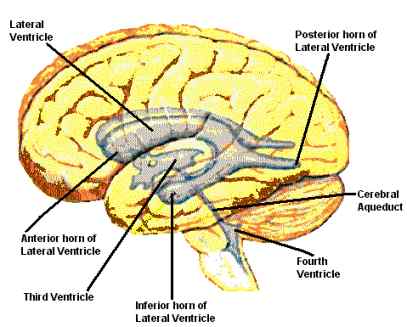
The third ventricle is a narrow, four-sided,
irregularly shaped opening in the middle of
the brain that provides a pathway for
cerebrospinal fluid. Cerebrospinal fluid is
the cushiony fluid that protects the brain
and spine from trauma. In addition to the
third ventricle, there is a fourth ventricle
and two lateral (side) ventricles, pictured to
the right. There is one lateral ventricle on
each side of the brain. Cerebrospinal fluid
is produced by a cluster of blood vessels
that line the ventricles. These clusters of
blood vessels are known as the choroid
plexus. Together, the ventricles of the brain
hold about a glassful of cerebrospinal fluid.
FEATURED BOOK: Neuroanatomy Through Clinical Cases (2nd ed.)
WHAT ARE THE DIFFERENT AREAS THAT SURROUND THE THIRD VENTRICLE?
As you can see from the picture above, the third ventricle is located below and between
the two lateral ventricles. The third ventricle has a narrow roof, a floor, and four walls.
The wall in the back is known as the posterior wall. The wall in the front is known as the
anterior wall. The walls on the side are known as the lateral walls.
"Where Medical Information is Easy to Understand"™
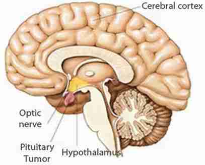
On each side of the third ventricle is a thalamus and the
hypothalamus. The thalamus is a pair of large oval structures that
sends out messages regarding sensation. The hypothalamus is an
area below the thalamus that is important for many bodily functions
such as sleep, appetite, and temperature control. For those who want
more specific detail, the structures that surround the third ventricle
are described below and divided by area:
FLOOR: The floor of the third ventricle is formed by the following
structures: the optic chiasm, mammillary bodies, tuber cinereum,
hypothalamus, subthalamus, infundibulum, posterior perforated
substance, and the upper part of the midbrain tegmentum.
The optic chiasm is an area in the brain where optic nerves (nerves that help us see) cross over. The
mammillary bodies are small, round, paired group of cells located in the bottom part of the hypothalamus.
You can see where the third ventricle, other ventricles, optic nerves, and hypothalamus are located in the
picture below.
The tuber cinereum is a bulge at the bottom of the
hypothalamus (see above). The subthalamus is an area
of the brain located between the hypothalamus and the
midbrain tegmentum (see below for description) that
processes information about vision and balance.
The infundibulum is a funnel-shaped stalk that extends
from the hypothalamus to the back part of the pituitary
gland. The pituitary gland is an area in the brain that
secretes (forms and gives off) chemicals known as
hormones, such as those that are important for growth.
Hormones are natural chemicals produced by the body
and released into the blood that have a specific effect
on tissues in the body.
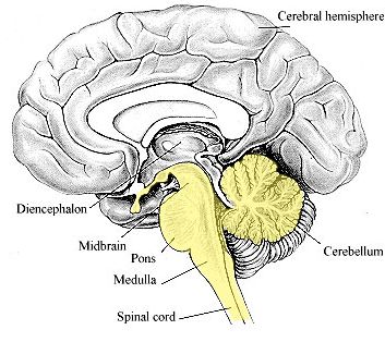
The posterior perforated substance is the bottom part of the interpeduncular fossa that allows plenty of room for branches of arteries (types of blood vessels that carry blood away from the heart) to pass through. The interpeduncular fossa is a deeply sunken in area of the midbrain. The midbrain is the top part of the brainstem. The brainstem is an area in the lower part of the brain that connects it with the spinal cord. The midbrain tegmentum is a term for the major part of the midbrain. The midbrain tegmentum is located in the top part of the midbrain.
Below is a picture of the midbrain and the rest of the brainstem (pons and medulla). You can see how the top of the midbrain forms part of the floor of the third ventricle. The third ventricle is located directly above the midbrain.
Below is a picture of the midbrain and the rest of the brainstem (pons and medulla). You can see how the top of the midbrain forms part of the floor of the third ventricle. The third ventricle is located directly above the midbrain.
ROOF: The narrow roof of the third ventricle is formed by
a thin membrane known as the tela choroidea. The tela
choroidea is a term for the combination of pia mater and
ependyma, which are both types of membranes. A
membrane is a thin layer of flexible tissue that covers
something.
Whereas ependyma is a type of membrane that lines the
ventricles of the brain, pia mater is a type of membrane
that lines other brain areas as well. The tela choroidea are
attached on each side to the tenia thalami. The tenia
thalami is the sharp edge or angle between the top and
middle surfaces of the thalamus (see picture).
ROOF: The narrow roof of the third ventricle is formed by a thin membrane known as the tela choroidea.
The tela choroidea is a term for the combination of pia mater and ependyma, which are both types of
membranes. A membrane is a thin layer of flexible tissue that covers something. Whereas ependyma is
a type of membrane that lines the ventricles of the brain, pia mater is a type of membrane that lines
other brain areas as well. The tela choroidea are attached on each side to the tenia thalami. The tenia
thalami is the sharp edge or angle between the top and middle surfaces of the thalamus (see picture).
WHAT ARE THE RECESSES OF THE THIRD VENTRICLE?
Recesses are small hollow areas or indentations. There are several recesses in the third ventricle:
PREOPTIC RECESS: This is located in the sharp angle between the bottom of the lamina terminalis and
the back of the optic chiasm. The lamina terminalis is a thin plate that passes upwards from the optic
chiasm (see picture). The optic chiasm is an area in the brain where optic nerves (nerves that help us
see) cross over.
INFUNDIBULAR RECESS: This funnel shaped area extends from the front part of the third ventricle into
the infundibulum (see previous section for description).
MAMMILLARY RECESS: This indentation is caused by the mammillary bodies (see picture) sticking into
the third ventricle. The mammillary recess is also known as the inframammillary recess.
PINEAL RECESS: From the back corner of the third ventricle, this recess extends into the stalk of the
pineal gland. See the last section for a description and picture of the pineal gland.
WHAT ARE SOME OTHER DETAILS ABOUT THE THIRD VENTRICLE?
The third ventricle is connected to each of the lateral ventricles and to the fourth ventricle. The area that
connects the third ventricle with each of the lateral ventricles is known as the interventricular foramen of
Monro (also known as the interventricular foramen and the foramen of Monro). The area that connects
the third ventricle with the fourth ventricle is known as the cerebral aqueduct (also known as the sylvian
aqueduct). The third ventricle extends from the lamina terminalis (see above) to the head of the cerebral
aqueduct.
WHAT ELSE IS THE THIRD VENTRICLE KNOWN AS?
The lateral ventricle is also known as the ventriculus tertius and the ventricle of diencephalon.
WHAT IS THE ORIGIN OF THE TERM, THIRD VENTRICLE?
Third ventricle comes from the Greek word "triotus" meaning "below second rank" and the Latin word
"venter" meaning "belly." Put the words together and you have "belly below second rank."