MedFriendly®


Magnetic Resonance Angiography
Magnetic resonance angiography (MRA) is a technique that
is used to provide extremely clear and detailed pictures of
blood vessels and the heart by using magnetic resonance
(see next question for description). The process of
obtaining pictures of the body by using magnetic resonance
is known as magnetic resonance imaging (MRI). Magnetic
resonance angiography is a type of magnetic resonance
imaging.
WHAT IS MAGNETIC RESONANCE?
Magnetic resonance is a complex phenomenon that
requires knowing the following:
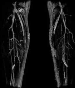
MRA of the legs.
FEATURED BOOK: MRA: Principles and Applications
Atom: The smallest part of an element that can exist alone or in combination with
something else. Atoms are so small that they cannot be seen even with high-powered
equipment.
Nucleus: The central structure of something, such as the central structure of an atom. A
nucleus is made up of particles called protons and neutrons.
Nuclei: More than one nucleus.
Magnetic field: The area around any magnet in which its effects can be detected.
"Where Medical Information is Easy to Understand"™
HOW IS MAGNETIC RESONANCE RELATED TO OBTAINING
PICTURES OF THE BODY?
Magnetic resonance is when the nuclei in atoms from certain
materials (when placed in a strong, constant, magnetic field), absorb
radio waves (a form of energy) sent from a source at certain
frequencies. In this case, frequency refers to the number of times
the radio waves move completely back and forth in one second.
Thus, radio waves can have high frequencies (moving back and
forth quickly) or low frequencies (moving back and forth slowly).
Before the nuclei are exposed to a magnetic field, the protons
(particles that make up the nuclei) are spinning in different
directions.
Exposing the nuclei to a magnetic field causes most of the protons to spin in such a way that they line up
next to each other in the same direction as the magnetic field.
At this point the atoms are in a state of low energy. Once the atoms are hit with short pulses of radio
waves and the radio waves are absorbed, the nuclei move from a state of low energy to a state of high
energy. In the state of high energy, the radio waves cause the protons to spin in such a way that they line
up in an opposite or different direction from the magnetic field. Thus, the radio waves briefly knock the
nuclei out of alignment (being lined up).
When the radio waves are turned off, the nuclei return to the state of low energy, go back to their natural
position, and give off photons (another form of energy). The photons produce energy signals which can be
analyzed by a receiver to produce very clear pictures of the body.
The energy signals are given off at the same frequency (see above for description) that the energy from
the radio waves was absorbed. It is mostly the energy emitted by hydrogen atoms that are analyzed by
MRI machines since the human body is made up mostly of water, and water is made of two parts
hydrogen (a nonmetallic element) and one part oxygen. In addition, each hydrogen atom contains one
proton that has a high likelihood of lining up in the same direction as the magnetic field.
The pictures of the body produced by using magnetic resonance are based on information that the
receiver in the MRI machine obtains about the strength and location of the signals sent out from the
nuclei. The pictures are so clear that they can detect very tiny changes in the body that may be the result
of a disease. In case you were wondering, resonance, in this case, means to cause a reaction (that is,
give off energy) by stimulating the movement of the parts inside an atom.
HOW DOES MAGNETIC RESONANCE ANGIOGRAPHY WORK?
The human body, like everything else, is made up of atoms. By applying the knowledge of magnetic
resonance (described above), scientists have discovered that the nuclei inside tissues that make up the
body respond differently than the nuclei of other tissues when placed in a constant, strong magnetic field
and are hit with radio waves. That is, the nuclei of some tissues send out signals (see last question for
more details) that last longer than those sent out from the nuclei of other tissues. For example, the signal
sent out from the nuclei of tumors (tissues that grow more rapidly than normal) lasts longer than the signal
sent out from the nuclei of normal tissue. This is why tumors show up so clearly on MRIs.
Scientists have figured out how to measure these signals to produce remarkably clear pictures of the
structures inside of the body. Special settings on the MRI machine are used to obtain pictures of the blood
vessels and/or the heart. Specifically, magnetic resonance angiography produces an image by increasing
the signal of flowing blood and decreasing the signal from other tissue. Pictures of tissue slices are
obtained that can be viewed from any angle or direction. Above is an example of a picture of the legs
using MRA.
WHY IS A MAGNETIC RESONANCE ANGIOGRAPHY PERFORMED?
Magnetic resonance angiographies are typically performed to detect, diagnose, and help treat disorders of
the heart and/or blood vessels. These disorders often involve a narrowing or blockage of arteries or
veins. Arteries are blood vessels that carry blood away from the heart, whereas veins are blood vessels
that carry blood to the heart. Magnetic resonance angiographies are also used to study stroke (a burst
artery or a blockage of an artery in the brain).
Magnetic resonance angiographies are commonly used to study the carotid arteries (located on both
sides of the neck) because they are common areas where blockages occur. They are also used to study
diseases of arteries in the head, kidneys, lung, legs, and the aorta (the largest artery in the body).
Magnetic resonance angiographies are also performed after treatment to find out if the arteries (or veins)
are still narrowed or blocked.
Magnetic resonance angiographies also provide pictures of veins (blood vessels that carry blood to the
heart). Magnetic resonance angiographies are also used to provide a picture of the size and thickness of
the heart. This allows the doctor to assess how much damage has been done to it by a heart attack or a
heart disease that has been occurring for a long time. Another use of magnetic resonance angiographies
is to detect aneurysms in patients that have a family history of this problem. An aneurysm is a weakening
of the wall of a blood vessel, which causes it to expand like a balloon and sometimes causes it to burst.
HOW IS A MAGNETIC RESONANCE ANGIOGRAPHY DIFFERENT FROM OTHER ANGIOGRAPHIES?
Traditional angiographies involve producing a picture of the inside structure of blood vessels and/or the
heart by using radiation (a type of energy) known as X-rays or gamma rays. Magnetic resonance
angiographies do not require the use of X-rays or gamma rays. Instead, magnetic resonance
angiographies use radio waves (another type of radiation). In addition, whereas traditional angiographies
are invasive, meaning that they require a needle, a wire, and a flexible, plastic tube to be inserted in the
blood vessels, magnetic resonance angiographies do not require this to be done. Thus, it is much more
convenient for patients to undergo a magnetic resonance angiography than a traditional angiography.
Traditional angiographies require the blood vessels to be filled with a substance (known as a contrast)
that radiation cannot pass through. This allows the blood vessels to be seen in contrast to the surrounding
areas that the x-rays can pass through. Magnetic resonance angiographies do not require the use of
contrast, but sometimes a special contrast is used in order to highlight an area of the body.
IS IT TRUE THAT MAGNETIC RESONANCE ANGIOGRAPHIES DO NOT USE RADIATION?
No. Magnetic resonance angiographies use radio waves, which are a form of radiation. They do not use
x-rays, however, which is another form of radiation that is more harmful to the human body than radio
waves.
IS A MAGNETIC RESONANCE ANGIOGRAPHY SAFE AND PAINLESS?
Yes to both. Magnetic resonance angiographies do not cause any pain and they are not known to cause
any type of tissue damage. The only time there would be pain is before the MRA, if a needle is used to
inject contrast material into a vein to highlight certain areas of the body.
WHAT DOES A MAGNETIC RESONANCE ANGIOGRAPHY MACHINE LOOK LIKE?
A typical magnetic resonance imaging machine looks like this:
Some people feel too confined in machines that resemble the picture because they are placed in a large
tube surrounded by a circular magnet. This concern led to the creation of newer MRI machines that do not
fully enclose the patient. Some of these newer machines are shorter, wider, and do not fully enclose the
patient. There are even some machines that are open on all sides. These newer MRI machines (known as
open MRIs) are being made increasingly available to people. The problem is that the pictures produced by
open MRI machines are not as clear as the pictures in traditional, closed MRI machines.
HOW SHOULD I PREPARE FOR A MAGNETIC RESONANCE ANGIOGRAPHY?
The first thing you want to do is to tell the person who does the magnetic resonance angiography if you
have any metal objects on or in your body. This is important because the procedure involves placing the
body in a magnetic field (see top of page for a description of this term), and will pull on anything containing
iron. Starting with objects on your body, you will need to remove hairpins, watches, eyeglasses, jewelry,
wigs that contain metal, removable dental work, and hearing aids. Tell the person who is doing the
magnetic resonance angiography if you have permanent red eyeliner or a red tattoo, because some red
dyes (colorings) have iron in it. Red dyes are rarely a problem, however.
Examples of some objects that may be inside your body for medical reasons are pins, screws, staples,
metal plates, joint replacements, and a pacemaker (an electrical device used to control heartbeat). If you
are going for a magnetic resonance angiography of the face or head, you should let it be known if you
have fillings in your teeth or braces because they can cause the pictures of these areas to look distorted.
You should also mention if you have a bullet inside of you from a past injury since these also contain
metal. You should even mention if you have worked with metals in the past. If there is any doubt whether
or not you have any metal inside of your body, you can have an x-ray to find out.
Be sure to mention what drug allergies you have and if you are pregnant. You can take your normal
medications before the exam. If you do not like being enclosed in a small space, you can be given
medication to help calm you down, or you can request to go in a machine that is more open (known as an
open MRI). Fewer than 1 in 20 patients are so nervous about being enclosed in a small space that they
need to take medication to help relax.
CAN I EAT OR DRINK BEFORE A MAGNETIC RESONANCE ANGIOGRAPHY?
In general, before undergoing a magnetic resonance angiography, most adults and young children are
allowed to eat or drink like they normally would. The exception is young children who are going to be given
a medication to relax them. These children should not eat or drink for about four hours before the exam.
Keep in mind that each place that performs magnetic resonance angiographies has different rules about
eating and drinking before the exam, so be sure to check with them.
WHAT HAPPENS ONCE I AM READY FOR THE MAGNETIC RESONANCE ANGIOGRAPHY?
Once the patient is ready, he/she may be asked to change into a hospital gown. The patient will then be
asked to sit down on a wheeled bed that moves him/her into a large tube surrounded by a circular magnet.
The patient lies down throughout the exam. There are several periods throughout the exam in which the
patient is told to stay still. At times, the magnet may be only a few inches from the patient's face. Some
loud knocking noises will be heard during some parts of the exam, which is due to switching the radio
waves on and off. The loud knocking noises are why some patients use earplugs during an MRI/MRA.
Most MRAs consist of 2 to 6 separate sequences (a succession of related pictures) in which pictures are
obtained. Each sequence takes between 2 to 15 minutes and provides pictures at different angles and
directions. The MRA can take between 10 and 60 minutes, depending on why it is being done.
As was mentioned earlier, it is possible that a material known as contrast will be administered to highlight
certain areas of the body. The contrast is administered by placing a needle in a vein. The contrast is then
injected into the vein through the needle during one of the imaging sequences. There may be some slight
pain where the needle is injected. The patient may feel a warm sensation in the area of the body being
studied shortly after the contrast is injected. This is a normal sensation, but it should be mentioned if it is
becoming a bother. Gadolinium is the name of the contrast used in MRAs and in MRI techniques in
general.
During the exam, the examiners leave the room and sit in a nearby room monitoring the process. The
patient can communicate with the examiner anytime over an intercom. Some places allow a friend to stay
nearby or a parent to stay nearby if a child is being examined. When the exam is finished, the patient will
be asked to wait just to make sure that no additional pictures are needed.
WHO INTERPRETS THE MAGNETIC RESONANCE ANGIOGRAPHY?
A type of doctor known as a radiologist, that has experience in interpreting MRIs, will analyze the results
and interpret the magnetic resonance angiography. He or she will then write a report about the results and
send it to your doctor. The radiologist may also discuss the results with your doctor.
HOW DO I FIND OUT THE RESULTS OF THE MAGNETIC RESONANCE ANGIOGRAPHY?
The results of the magnetic resonance angiography will be provided to you by the doctor that referred you
to get the test. This process can be speeded up if the MRI center sends the results and images to your
doctor over the Internet.
WHAT ARE THE BENEFITS OF MAGNETIC RESONANCE ANGIOGRAPHY?
1. An MRA provides very clear pictures of blood vessels and the heart.
2. Unlike traditional angiographies, there is no risk of harming arteries because a catheter (a flexible,
plastic tube) is not used. In traditional angiographies, a catheter is pushed through the arteries.
3. An MRA takes less time and costs less money than a traditional angiography.
4. The recovery time for an MRA is less when compared to the recovery time for a traditional
angiography.
5. Surgery can often be avoided based on the results of the MRA. If surgery is needed it can be done
more accurately because the surgeon will use the pictures from the MRA as a guide.
6. When compared to another imaging technique known as a Computerized Axial Tomography (CT) scan,
an MRA does not expose patients to x-rays and requires patients to stay still for shorter periods of time.
7. MRAs do not require the use of contrast, unlike traditional angiographies and CT scans. MRAs can still
provide clear pictures of the blood vessels and heart without using contrast.
8. The contrast material used in an MRA does not contain iodine (a nonmetallic element), unlike the
contrast material used in tradition angiographies and in CT scans. Iodine leads to a risk of kidney damage
and allergic reactions. The chances of having an allergic reaction to the contrast used in angiographies
and CT scans are low, but allergic reactions to the contrast used in MRAs are even lower.
WHAT ARE SOME PROBLEMS WITH MAGNETIC RESONANCE ANGIOGRAPHY?
1. Some people cannot have the procedure done because they get extremely nervous when they are
inside of enclosed spaces.
2. If a patient requires medication to relax during an MRA, a nurse needs to be there to monitor the
patient.
3. Women should not have an MRA during the first three months of pregnancy because the effects of this
procedure on the developing baby are unknown. The only exception is when there is a major risk in
missing the correct diagnosis by not doing the procedure. An alternative procedure is an ultrasound, which
uses types of sound waves to produce images of the body.
4. Women who are breast-feeding their babies, and need to receive contrast, should ask their doctor what
to do. Sometimes, the breast milk is pumped out before the MRA so it can be given to the baby until the
contrast leaves the woman's body.
5. Any metal objects in the body that are not detected before the MRA is done may be negatively affected
by the procedure. If the metal object, such as a pacemaker (an electrical device used to control
heartbeat), is near the area of the body that needs to be studied, the resulting pictures may be distorted.
6. An MRA should not be done in patients with bullet fragments in the body, metal objects in the eye, metal
objects placed in the ear, a pacemaker, an artificial heart valve (a structure that opens and closes to
influence blood flow), or a neurostimulator in the body. A neurostimulator is a device that excites a part of
the nervous system in the body, such as the brain or spinal cord.
7. Although the pictures produced by MRAs are clear, they are not as clear as the pictures from traditional
angiographies.
8. It may sometimes be difficult to tell the difference between arteries and veins on an MRA.
9. Unlike CT scans (see last question), MRAs do not produce pictures of calcium (a metallic element).
WHAT IS THE ORIGIN OF THE TERM, MAGNETIC RESONANCE ANGIOGRAPHY?
Magnetic comes from the Greek word "magnesia." Magnesia refers to "Magnes lithos" which is a rock in
Magnesia. Magnesia is a region in Thessaly, Greece. It is here where ore (a mineral containing something
valuable) was found that had the property of a magnet. Resonance comes from the Latin word "resonare"
meaning "to sound again." Angiography comes from the Greek word, "angeion" meaning "a vessel or
opening in the body," and the Greek word "graphe" meaning "to write." Put the two words together and you
get "to write (about) a vessel or opening of the body." The word "angeion" comes from the Greek word
"angos" meaning "a vessel or bucket," and the Greek word "eion" meaning "small."