


MedFriendly®


Magnetic Resonance
In order to describe such a complex topic as magnetic resonance
in an easy to understand manner, some basic terms need to be
explained. If you already know these terms, skip to the next
section.
Atom: The smallest part of an element that can exist alone or in
combination with something else. Atoms are so small that they
cannot be seen even with high-powered equipment.
FEATURED BOOK: MRI: Physical and Biological Principles
Nucleus: The central structure of something, such as the central structure of an atom. A
nucleus is made up of particles called protons and neutrons.
Nuclei: More than one nucleus.
Magnetic field: The area around any magnet in which its effects can be detected.
KNOWING THE TERMS ABOVE, WHAT IS MAGNETIC RESONANCE?
Magnetic resonance is when the nuclei in atoms from certain materials (when placed in a
strong, constant, magnetic field), absorb radio waves (a form of energy) sent from a
source at certain frequencies. In this case, frequency refers to the number of times the
radio waves move completely back and forth in one second. Thus, radio waves can have
high frequencies (moving back and forth quickly) or low frequencies (moving back and
forth slowly).
"Where Medical Information is Easy to Understand"™
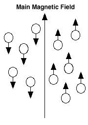
Before the nuclei are exposed to a magnetic field, the protons
(particles that make up the nuclei) are spinning in different
directions. Exposing the nuclei to a magnetic field causes most of
the protons to spin in such a way that they line up next to each
other in the same direction as the magnetic field. This causes the
nuclei to look like they do to the right. As you can see above, the
nuclei are lined up next to each other. At this point the atoms are in
a state of low energy.
ONCE THE NUCLEI ARE HIT WITH RADIO WAVES, WHAT
HAPPENS NEXT?
Once the atoms are hit with short pulses of radio waves and are absorbed, the nuclei move from a state
of low energy to a state of high energy. In the state of high energy, the radio waves cause the protons to
spin in such a way that they line up in an opposite or different direction from the magnetic field. Thus, the
radio waves briefly knock the nuclei out of alignment (being lined up).
When the radio waves are turned off, the nuclei return to the state of low energy, go back to their natural
position, and give off photons (another form of energy). The photons produce energy signals which can
be analyzed by a receiver in a magnetic resonance imaging (MRI) machine to produce very clear pictures
of the body, such as this picture of the brain:

The energy signals are given off at the same frequency (see last question for description) that the energy from the radio waves was absorbed. It is mostly the energy emitted by hydrogen atoms that are analyzed by MRI machines since the human body is made up mostly of water, and water is made of two parts hydrogen (a nonmetallic element) and one part oxygen. In addition, each hydrogen atom contains one proton that has a high likelihood of lining up in the same direction as the magnetic field. The pictures of the body produced by using magnetic resonance are based on information that the receiver in the MRI machine obtains about the strength and location of the signals sent out from the nuclei. The pictures are so clear that they can detect very tiny changes in the body that may be the result of a disease. In case you were wondering, resonance, in this case, means to cause a reaction (that is, give off energy) by stimulating the movement of the parts inside an atom.
HOW IS MAGNETIC RESONANCE HELPFUL TO HUMANS?
The human body, like everything else, is made up of atoms. By applying the knowledge of magnetic
resonance, scientists have discovered that the nuclei inside certain tissues of the body respond
differently than the nuclei of other tissues when placed in a constant, strong magnetic field and are hit with
radio waves.
That is, the nuclei of some tissues send out signals (see last question for more details) that last longer
than those sent out from the nuclei of other tissues. For example, the signal sent out from the nuclei of
tumors (tissues that grow more rapidly than normal) lasts longer than the signal sent out from the nuclei of
normal tissue. This is why tumors show up so clearly on MRIs.
Scientists have figured out how to measure these signals to produce remarkably clear pictures of the
structures inside of the body. As was mentioned before, this technique is known as magnetic resonance
imaging. With an MRI, pictures of tissue slices are obtained that can be viewed from any angle or
direction. A special type of MRI is known as an MRA (magnetic resonance angiography), which provides
extremely clear and detailed pictures of blood vessels and the heart by increasing the signal of flowing
blood and decreasing the signal from other tissue.
WHO DISCOVERED MAGNETIC RESONANCE AND WHEN?
Magnetic resonance was discovered in 1946 by Felix Bloch and Edward Purcell. They made their
discoveries separate from one another and each won the Nobel Prize in 1952.
WHAT ELSE IS MAGNETIC RESONANCE KNOWN AS?
Magnetic resonance is also known as nuclear magnetic resonance.
WHAT IS THE ORIGIN OF THE TERM, MAGNETIC RESONANCE?
Magnetic comes from the Greek word "magnesia." Magnesia refers to "Magnes lithos" which is a rock in
Magnesia. Magnesia is a region in Thessaly, Greece. It is here where ore (a mineral containing something
valuable) was found that had the property of a magnet. Resonance comes from the Latin word "resonare"
meaning "to sound again."
