MedFriendly®


Bronchocele
A bronchocele is a part of the bronchus that is filled with
mucous and which is completely enclosed so that the
mucous has no way to drain out. A bronchus is a type
of small airway connected to the lungs. The part of the
bronchus in a bronchocele is typically widened.
Bronchoceles can be caused by obstructions such as
scarring, a tumor, a stricture (narrowing of a tube-
shaped structure), presence of foreign body, or
abnormal closure from birth (known as congenital
atresia). Tumors are abnormal masses of tissue that
form when cells in a certain area of the body reproduce
at an increased rate. This is a rare cause of a
bronchocele but when it happens it is usually caused by
a carcinoma.
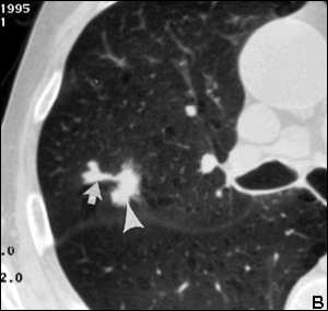
A bronchocele shown on imaging.
FEATURED BOOK: Muller's Diseases of the Lungs
A carcinoma is any malignant cancer that arises from cells in the covering surface layer
or membrane (outer covering) that lines an organ of the body. Cancer is any of a large
group of malignant diseases characterized by an abnormal, uncontrolled growth of new
cells in one of the body organs or tissues. Another type of tumor that can cause a
bronchocele is an adenoma, which is a benign (non-cancerous) tumor of a gland.
A non-obstructive cause of a bronchocele is asthma because people with asthma are
predisposed to infection from aspergillus (a type of mold). This is known as allergic
bronchopulmonary aspergillosis.
"Where Medical Information is Easy to Understand"™
When the body produces too much mucous, this can lead to
formation of a bronchocele, such as in asthma and cystic fibrosis.
Cystic fibrosis is a disease passed down through families that
causes thick mucous to build up in the lungs, digestive tract, and
other areas of the body. Another cause of a bronchocele is
tuberculosis (a type of lung infection).
shortness of breath, cough, chest pain, fatigue, spitting up mucous,
and coughing up blood or blood-stained products. In some cases,
there may be no signs or symptoms.
Bronchoceles can be detected via a chest x-ray but is best diagnosed via a CT scan of the chest. CT
(computerized tomography) scanning is an advanced imaging technique that uses x-rays and computer
technology to produce more clear and detailed pictures than a traditional x-ray. These diagnostic imaging
techniques can reveal multiple bronchoceles in a single patient. Bronchoceles are usually bigger than one
centimeter and will appear to have branching fingers on an x-ray. Both the chest x-ray and the CT may
show that air has been trapped in the region of the bronchocele. If the lung collapses due to obstruction,
an x-ray will be unable to identify the bronchocele but a CT scan will still be able to detect it.
Bronchoceles can be managed by treating the underlying cause (e.g., antibiotics). Physical therapy may
also be a part of treatment. In some cases, surgical removal of the obstruction may be needed. This can
be done via a bronchoscopy, in which a device is passed through the mouth or nose, through the
windpipe, and into the lungs. During this procedure, a biopsy can be performed. A biopsy is the process of
removing living tissue or cells from organs or other body parts of patients for examination under a
microscope or in a culture to help make a diagnosis, follow the course of a disease, or estimate a
prognosis. A culture is an artificial way to grow cells or tissues in the laboratory.
If mucous drainage in the bronchus is not obstructed, the condition is referred to as a mucoid impaction of
bronchus. Bronchocele comes from the Greek word bronchus meaning windpipe, and the Latin word
kele meaning hernia. Put the words together and you get windpipe hernia.