


MedFriendly®


Lateral Ventricles
The lateral ventricles are two curved
openings (shaped like a horseshoe) located
deep within the top section of the brain, that
provide a pathway for cerebrospinal fluid.
Cerebrospinal fluid is the cushiony fluid that
protects the brain and spine from trauma.
There is one lateral ventricle on each side of
the brain. In addition to the two lateral
ventricles, there is the third ventricle and the
fourth ventricle, both of which are connected
by the cerebral aqueduct. The two lateral
ventricles are the largest of all the ventricles
in the brain. They are surrounded by
periventricular white matter. White matter is
a group of white nerve fibers that conduct
nerve impulses quickly.
FEATURED BOOK: Neuroanatomy Through Clinical Cases (2nd ed.)
Cerebrospinal fluid is produced by a cluster of blood vessels that line the ventricles.
These clusters of blood vessels are known as the choroid plexus. Together, the
ventricles of the brain hold about a glassful of cerebrospinal fluid.
WHAT ARE THE DIFFERENT PARTS OF THE LATERAL VENTRICLE AND WHAT
STRUCTURES SURROUND THEM?
"Where Medical Information is Easy to Understand"™
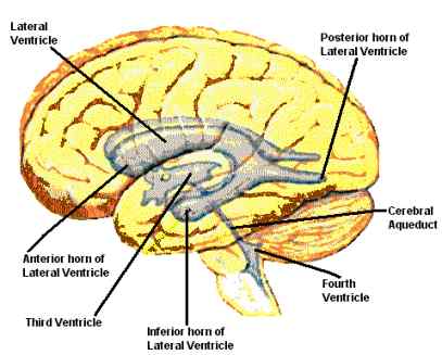
Each lateral ventricle consists of several parts, pictured above. One part is called the anterior (frontal)
horn of the lateral ventricle, which is the front part of the lateral ventricle. The anterior horn is located in
the front section of the brain, known as the frontal lobe. The anterior horn is positioned in front of the
interventricular foramen, which connects the lateral ventricles to the third ventricle.
Above the anterior horn of the lateral ventricle is the corpus callosum (also pictured above). The corpus
callosum is a group of nerve fibers that connect both sides of the brain and allow them to communicate.
The anterior horns of the lateral ventricle are separated by two triangular-shaped membranes known as
the septum pellucidum. Directly below the anterior horn of the lateral ventricle is the head of the caudate
nucleus. The caudate nucleus is a paired group of nerve cells that play an important role in body
movements.
The back of the lateral ventricle is known as the posterior horn of
the lateral ventricle. This is sometimes referred to as the occipital
horn of the lateral ventricle because it extends into a section of the
back part of the brain called the occipital lobe. More specifically,
each posterior horn extends into the white matter of the occipital
lobe. White matter is a group of white nerve fibers that conduct
nerve impulses quickly. Like the anterior horn of the lateral ventricle,
the corpus callosum is also above the posterior horn.
The center part of the lateral ventricle is appropriately named the
central part of the lateral ventricle. The central part of the lateral
ventricle is also known as the central body and the cella media.
Above the central part of the lateral ventricle is the corpus callosum (see above). The middle of the
central part of the lateral ventricle is formed by the back part of the septum pellucidum (see above).
Below the central part of the lateral ventricle are parts of the caudate nucleus (see above), parts of the
choroid plexus (the blood vessels in the lateral ventricle that produce cerebrospinal fluid), the thalamus,
and the fornix cerebri. The thalamus is a pair of large oval structures that sends out messages regarding
sensation. The fornix cerebri is an archlike body of nerve fibers that projects outwards from the
hippocampus (a structure in the lower part of the brain that is important for memory).
The bottom of the lateral ventricle is known as the inferior horn of the lateral ventricle. This is sometimes
referred to as the temporal horn of the lateral ventricle because it is in an area of the brain called the
temporal lobe. Above the inferior horn is white matter of the brain. The middle of the inferior horn is formed
by the tail of the caudate nucleus (see above) and the stria terminalis. The stria terminalis is a thin bundle
of fibers that forms a pathway in the limbic system. The limbic system is an area near the edge of the
middle part of the brain that is important for producing emotions.
The amygdala bulges into the end of the inferior horn. The amygdala is a round mass of gray matter (a
gray appearing substance) in the temporal lobe. The temporal lobes are located on the sides of the brain
by the ears. The bottom and middle part of the internal horn is formed by the hippocampus (see above),
fimbria of the hippocampus, and collateral eminence. The fimbria of the hippocampus is a band of fibers
alongside the hippocampus. The collateral eminence is a bulging of white matter that is found near the
hippocampus.
WHAT ARE SOME OTHER DETAILS ABOUT THE LATERAL VENTRICLES?
The lateral ventricles take on a curved shape that conforms to the shape of each half of the brain. The
lateral ventricles pass through all four lobes (sections) of the brain. As you can see from the pictures
above, the lateral ventricles are connected to the third ventricle. The area that connects the lateral
ventricles with the third ventricle is known as the interventricular foramen of Monro (also known as the
interventricular foramen and the foramen of Monro). The choroid plexus, which are the blood vessels in
the lateral ventricle that produce cerebrospinal fluid, are found in the central part of the lateral ventral and
the inferior horn, but not in the anterior or posterior horns.
WHAT ELSE ARE THE LATERAL VENTRICLES KNOWN AS?
The lateral ventricle is also known as the ventriculus lateralis and the ventricle of cerebral hemisphere.
WHAT IS THE ORIGIN OF THE TERM, LATERAL VENTRICLES?
Lateral ventricles comes from the Latin word "latus" meaning "side" and the Latin word "venter" meaning
"belly." Put the words together and you have "side belly."