MedFriendly®


Syringomyelia
Syringomyelia is a rare condition in which long, fluid filled spaces are
present in the central gray matter (a gray looking substance) of the
spinal cord and surrounded by thick tissues made of glial cells. Glial
cells maintain and support other cells. A cell is the smallest, most
basic unit of life, that is capable of existing by itself. Because
syringomyelia usually gets worse over time it is known as a
progressive illness.
WHAT CAUSES SYRINGOMYELIA?
In syringomyelia, cerebrospinal fluid enters the spinal cord an forms
an opening known as a syrinx. Cerebrospinal fluid is a cushiony fluid
that protects the brain and spine.
FEATURED BOOK: Syringomyelia: A Disorder of CSF Circulation
Over time, the opening gets bigger and longer, destroying the center of the spinal cord.
Nerve fibers inside the spine can also get damaged. There are many possible events that
can lead to syringomyelia. About 70% of syringomyelia cases are due to abnormalities in
the back of the brain, such as Chiari malformation. Chiari malformation is a condition in
which important parts of the back, bottom part of the brain extends downwards through
the bottom of the skull where the brain joins the spinal cord.
The brain structures that extend downwards in Chiari's malformation are the cerebellar
tonsils, the pons, and the medulla oblongata. The cerebellar tonsils are two structures at
the bottom part of the cerebellum.
"Where Medical Information is Easy to Understand"™
The cerebellum is an area in the back, bottom part of the brain that
is plays an important role in movement and coordination. The pons
is very important for sleep and arousal and the medulla oblongata is
extremely important for controlling breathing. In Chiari's
malformation, the cerebellar tonsils, pons, and medulla oblongata
stick out because there is not enough room at the back of the brain.
Normally, the cerebellum, pons, and medulla oblongata rest in an
indented area of bone in the lower, back part of the skull known as
the posterior cranial fossa. However, the posterior cranial fossa is
abnormally formed in people with Chiari's malformation, providing
less space for these important brain areas. With less space to be
in, these areas push downwards into the spinal cord.
Syringomyelia can be due to a tumor or trauma to the spinal cord. It can also be due to a tethered spinal
cord, in which the spinal cord is stretched because it is abnormally fastened to an immovable structure.
Meningitis, which is an inflammation of the meninges (the three outer coverings of the brain and spinal
cord), can lead to syringomyelia as well. The middle of these coverings, known as the arachnoid
membrane, can also get inflamed and lead to syringomyelia. The events mentioned above cause or can
cause damage to the spinal cord. It is this damage that can cause the fluid-filled opening to form. The fluid-
filled opening will form in the area of the damage. In some cases, the fluid-filled cavity does not form until
years after the spinal cord is injured.
Only rarely is syringomyelia due to hemorrhage (loss of a large amount of blood in a short period of time).
Some people with syringomyelia have low-grade astrocytomas (a type of brain tumor) or abnormal
formations of blood vessels in the spine.
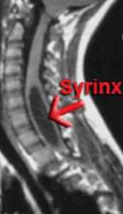
WHAT DOES SYRINGOMYELIA LOOK LIKE?
The long spaces in syringomyelia look like tubes in the spinal cord. It can also look like one big tube in the
spinal cord. Here is an example of what syringomyelia looks like in this scan of the spine:
WHAT ARE THE SIGNS AND SYMPTOMS OF SYRINGOMYELIA?
The signs and symptoms of syringomyelia are numerous and will differ depending on the size of the fluid-
filled spaces and where they are located. People with syringomyelia generally experience long-term pain,
headaches, and paresthesias (abnormal sensations). This is usually followed by wasting away of the
muscles in the hands, muscle weakness, and a loss of pain and temperature sensations in the hands and
arms. The sense of touch, however, remains unchanged. Painless lesions (abnormal changes) follow, as
does spastic paralysis in the legs. Spastic paralysis is a loss of muscle function in addition to contractions
or shortening of the muscles.
Scoliosis of the lumbar (lower) spine is also common in syringomyelia. Scoliosis is a condition in which
the spine curves abnormally to the side. Another common sign is losing control of the ability to poop and
pee. Osteoporosis, which is an abnormal loss of bone thickness and a wearing away of bone tissue, is
also common in syrigomyelia.
The signs and symptoms of syringomyelia usually begin early in adulthood, are more common in males,
and usually involve the cervical spine (the part of the spine by the neck). The signs and symptoms of
syringomyelia tend to develop slowly but can occur suddenly. Sudden onset of symptoms due to
syringomyelia is usually accompanied by coughing and straining.
HOW IS SYRINGOMYELIA TREATED?
The only treatment for syringomyelia is surgery. However, surgery is only recommended when the
condition is severe enough. When this is the case, the first step is to find a neurosurgeon that specializes
in syringomyelia. A neurosurgeon is a doctor that operates on the spine, brain, and/or nerves outside the
brain and spine.
The surgery is done to correct the condition that caused the syringomyelia. For example, if syringomyelia
is due to Chiari's malformation (see earlier for description), the goal is to provide more space for the
cerebellum in the back of the skull and in the upper neck area. By doing this, the flow of cerebrospinal fluid
returns to normal. This causes the fluid-filled space that is characteristic of syringomyelia to flatten out or
disappear.
In some cases, to get rid of the fluid-filled space, a flexible tube called a shunt can be placed inside of it.
From here, the tube will drain the extra fluid into the space between layers that line the belly. The fluid will
then be absorbed along the wall of the belly. If surgery succeeds, it will usually stop the condition from
getting worse and possibly help to moderately decrease the symptoms. Unfortunately, over time, surgery
is not always successful and more than one surgery may be needed. In cases in which surgery is not
needed, the patient needs to be monitored often by the treating doctor with physical examinations and
scans of the spine.
HOW MANY PEOPLE HAVE SYRINGOMYELIA?
In the United States, it is estimated that about 1 in 18,000 people have syringomyelia. In total, it is
estimated that about 21,000 people in the United States have syringomyelia. Both of these figures are
likely underestimates, however, because scientists have not studied this issue closely enough yet.
WHEN WAS SYRINGOMYELIA FIRST DESCRIBED?
Syringomyelia was first described over 400 years ago.
WHAT ELSE IS SYRINGOMYELIA KNOWN AS?
Syringomyelia is also known as Morvan's disease, myelosyringosis, syringomyelus, syringomyelic
syndrome, and hydrosyringomyelia.
WHAT IS THE ORIGIN OF THE TERM, SYRINGOMYELIA?
Syringomyelia comes from the Greek word "syrinx" meaning "tube," and the Greek word "myelos" meaning
"marrow." Put the two words together and you get "tube(s) (in the) marrow."