


MedFriendly®


Ultrasound Scanning
Ultrasound scanning is a procedure that uses high-
frequency sound waves to produce images of internal
body structures. The frequency (or pitch) of the sound
waves are so high that they cannot be heard by the
human ear.
HOW DOES ULTRASOUND SCANNING WORK?
After the patient lies down on an examination table, a
clear, cool, jelly-like substance is squirted over the area
of the body to be examined. This jelly-like substance can
easily be dissolved in water.
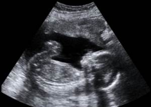
Ultrasound showing a fetus.
After the gel is applied, a wand-like object (known as a transducer) is gently rubbed on
the skin that covers the body part to be examined. The transducer emits high frequency
sound waves into the tissues of the body being examined.
The sound waves bounce off the internal body parts and reflect back as an echo to a
computer that the transducer is attached to. The echo is converted into electrical
impulses by the computer and translated into moving images of the body part being
examined. The moving images are recorded on videotape for analysis. Some images will
appear black when they do not produce echoes (known as anechoic) whereas others will
appear various shades of gray. As another example of how ultrasound scanning detects
abnormalities, solid masses (such as tumors), give off strong echoes and show up bright
on ultrasound images. Tumors are abnormal masses of tissue that form when cells in a
certain area of the body reproduce at an increased rate.
In some cases, the transducer may need to be inserted inside the vagina or rectum. The
former allows for a more detailed inspection of the uterus and fetus (an unborn living
being), especially during the very early stages of pregnancy. The latter is very helpful for
assessing the prostate gland. The prostate gland is a organ near the bladder that
produces a fluid that is part of semen (male repoductive fluid).
"Where Medical Information is Easy to Understand"™
WHY IS ULTRASOUND SCANNING USED?
Ultrasound scanning is used for assessment,
diagnosis, and screening of numerous potential
abnormalities. It is usually performed as an
outpatient procedure. As an example of
assessment, ultrasound scanning is performed
periodically during pregnancy to determine whether
normal development is occurring. Multiple
pregnancies can be detected with ultrasound
scanning. Ultrasound scanning is used to determine
the position, size, and age of the fetus.

Ultrasound scanning is also used to determine the location of the placenta. The placenta is an organ in
a female that nourishes a baby during pregnancy.
If an ultrasound is being performed in the early stage of a womans pregnancy, she will need to drink
several glasses of water about an hour before the scan. The reason for this is to make the bladder full
because this allows the uterus and fetus to be seen better. The uterus is a hollow organ in a female's
body where the egg is implanted and the baby develops. The woman will also be asked not to urinate for
about an hour before the scan.
As an example of how the technique aids in diagnosis, ultrasound scanning of breast tissue is often
performed to determine if there are any abnormalities such as cancer or cysts. A cyst is an abnormal
lump, swelling, or sac that contains fluid, a part solid material, or a gas, and is covered with a
membrane. A membrane is a thin layer of flexible tissue that covers something. Ultrasound scanning can
be used as a guide to assist in outpatient procedures such as the insertion of small catheters to drain
infections. A catheter is a flexible, hollow tube that is inserted into an opening or blood vessel in the
body, with the main purpose of allowing fluid to pass from or into these areas.
Ultrasound scanning can also be used as a guide to help with biopsies of tumors which are done by
inserting a thin needle into them. A biopsy is the process of removing living tissue or cells from organs
or other body parts of patients for examination under a microscope or in a culture to help make a
diagnosis, follow the course of a disease, or estimate a prognosis.
WHAT BODY PARTS ARE COMMONLY ASSESSED WITH ULTRASOUND SCANNING?
In children and adults, body parts commonly examined with ultrasound scanning include the spleen,
kidneys, and liver. The spleen is an organ next to the stomach that helps fight infection and removes
and destroys worn-out red blood cells. Red blood cells are cells that help carry oxygen in the blood. The
kidneys are two organs located on each side of the spine, behind the stomach. The kidneys filter
(remove) wastes from the blood. The liver is the largest organ in the body and is responsible for filtering
harmful chemical substances, producing important chemicals for the body, and other important
functions.
In females, the pelvis is commonly examined with ultrasound scanning. The pelvis is a massive bone
made of hip bones on each side and the front, while the back part is made of the sacrum and the
coccyx (also known as the tailbone). The sacrum is a large triangle shaped bone in the lower part of the
spine. The coccyx is a beaked shaped bone that makes up part of the back of the pelvis.
As opposed to children, adults are more likely to have the following body parts assessed with ultrasound
scanning: breasts, testicles, uterus, prostate, thyroid, gallbladder, ovaries, parathyroid glands, arteries,
and veins. A description of some of the more unfamiliar body parts follows. The thyroid gland is a
butterfly-shaped organ located in front of the neck that produces a natural chemical known as hormones
that affect virtually every cell in the body and many functions such as disease fighting, heart rate,
energy level, and skin condition.
The gallbladder is a small, pear shaped sac, located under the liver, which helps store and transport bile
to the first part of the small intestine (known as the duodenum). Bile is a bitter, yellow-green substance
released from the liver that carries away waste products. The small intestine is the part of the digestive
system that takes in all of the nutrients (healthy substances) that the body needs. Ovaries are organs in
the female body that normally produce eggs.
The paratrhyroid glands are four small glands located on either side of the thyroid gland which regulate
the level of calcium in the blood. Arteries are blood vessels that carry blood away from the heart. Veins
are blood vessels that carry blood to the heart. Assessing whether arteries that supply blood to the
brain are too narrow is a way ultrasound scanning is used to determine the risk of developing stroke. A
stroke is a burst artery or a blockage of an artery in the brain.
IS THERE RADIATION IN ULTRASOUND SCANNING?
No. Ultrasound scanning does not involve exposure to radiation, like x-rays do. For this reason, the
technique is considered very safe and is why it is often used to assess pregnant women.
IS ULTRASOUND SCANNING PAINFUL?
No. Ultrasound scanning is essentially pain-free.
IS ANY SPECIAL CARE REQUIRED AFTER ULTRASOUND SCANNING?
No. Ultrasound scanning does not require any special care afterwards.
WHO ACTUALLY PERFORMS ULTRASOUND SCANNING?
Ultrasound scanning is usually performed by a healthcare professional known as a sonographer.
Sonographers are specifically trained in ultrasound scanning. Physicians who are properly trained can
also perform ultrasounds. There are special training schools available to teach people to become a
diagnostic medical ultrasound technolgist.
WHAT ELSE IS ULTRASOUND SCANNING KNOWN AS?
Ultrasound scanning is also known as sonography, echography, and ultrasonography.
WHAT IS THE ORIGIN OF THE TERM, ULTRASOUND?
Ultrasound comes from the Latin word "ultra" meaning "beyond," and the Latin word "sonus" meaning
"sound." Put the words together and you get "beyond sound."