MedFriendly®


Atrial Septal Defect
To understand what an atrial septal defect is,
it is first necessary to know some basic
information about the heart. The heart is
divided into two halves by a wall known as
the septum. Each half is divided into an upper
and lower chamber. The chambers on the
bottom of the heart are called ventricles.
Each chamber on the top of the heart is
called an atrium. More than one atrium is
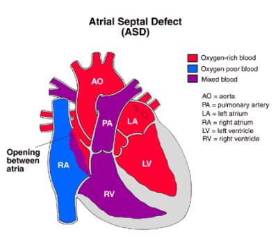
known as atria. An atrial septal defect is type
of birth defect characterized by an abnormal
opening somewhere in the wall (septum) that
divides the left and right atrium. An atrial
septal defect is one of the most common
heart defects seen and it is the most common
birth defect of the heart seen in adults.
FEATURED BOOK: Clinical Cardiology Made Ridiculously Simple
WHAT DO THE VENTRICLES AND ATRIA OF THE HEART DO?
The atria are the chambers of the heart that receive blood and the ventricles are the
chambers of the heart that pump blood out of the heart. The left atrium receives blood
with a high level of oxygen from the lungs. The left ventricle pumps out blood with the high
level of oxygen to the body. The left atrium sends blood to the left ventricle and the right
atrium sends blood to the right ventricle. The right atrium receives blood with a low level
of oxygen from the body. The right ventricle pumps out blood with the low level of oxygen
to the lungs. The lungs are two organs in the body that help people breathe.
"Where Medical Information is Easy to Understand"™
Because there are so many possible causes of arthralgia,
treatment is based on addressing the cause (e.g., treating an
infection with antibiotics, stopping a medication causing an allergic
reaction). Severe cases may require removal (i.e., arthrectomy)
and replacement of a joint through surgery. Medications to reduce
inflammation can treat inflammatory causes of joint pain. In
addition to treating the underlying cause, other treatment can
include pain medication, stretching exercises, and application of
hot and/or cold compresses. To learn more about the science of
joints, see the entry of arthrology. Arthralgic and arthrodynic
means pertaining to or affected with arthralgia. Arthralgia is also
known as arthrodynia.
ARE THERE DIFFERENT TYPES OF ATRIAL SEPTAL DEFECTS?
Yes. To understand the 3 main types of atrial septal defects it is important to know the following
information. The undeveloped child has normal openings in the wall of the atrium for a period of time.
These openings are supposed to close up and not be present when the child is born. In the fifth week of
pregnancy, the presence of the first hole can be seen in the unborn child's heart. This hole is known as
the ostium primum or the foramen primum (which means "first hole"). The ostium primum is located in the
lower part of the wall that divides the right and left atrium.
In the sixth week of pregnancy, the presence of the second hole can be seen in the unborn child's heart.
This hole is known as the ostium secundum or the foramen secundum (which means "second hole"). The
ostium secundum is located in the upper part of the wall that divides the right and left atrium.
Atrial septal defects are classified into three main types:
1. OSTIUM PRIMUM DEFECT: Normally, the first septum (wall) of the atrium will fuse with the endocardial
cushions. Endocardial cushions are a pair of thickened sections of tissue in the atrium of an unborn child.
In an ostium primum defect, there is inadequate development of the endocardial cushions. As a result,
they do not fuse with the first septum (wall) of the atrium and a hole develops in the lower part of that wall.
2. OSTIUM SECUNDUM DEFECT: In this type, the opening in the second septum (wall) of the heart of the
unborn child fails to fuse with the endocardial cushions. The hole that develops is usually 1 to 4
centimeters. 60% of atrial septal defects are ostium secundum defects.
3. SINUS VENOUS DEFECT: In this type, the top portion of the atrium fails to develop. This type of atrial
septal defect is associated with abnormal drainage into the right atrium from the vein in the upper right
lobe (part) of the lungs. Veins are blood vessels that carry blood to the heart. 10% of atrial septal defects
are sinus venous defects.
HOW SEVERE IS AN ATRIAL SEPTAL DEFECT?
The severity of an atrial septal defect depends on the size and location of the hole in the wall of the
atrium. A small hole in the wall of the atrium would be less severe than a large hole. Some atrial septum
defects are the size of a pinhole whereas others are characterized by a total absence of the wall that
divides the left and right atrium.
The size and location of the hole is related to when the development of the wall of the atrium stopped
when the person was an embryo. With regards to humans, an embryo is a very early form of the person
from about 2 weeks after it was conceived until the end of the 7th or 8th week. Major organ systems (such
as the heart) develop during the embryonic (embryo) stage.
CAN ATRIAL SEPTAL DEFECTS BE INHERITED?
Yes. There is strong evidence in some cases that atrial septal defects can be inherited from one's
parents.
WHAT ARE THE CONSEQUENCES OF AN ATRIAL SEPTAL DEFECT?
The consequences of an atrial septal defect depend on the size of the hole. Smaller holes sometimes
close by themselves and usually do not cause any problems to the child. Some children may experience
frequent respiratory infections. It is possible not to experience any signs or symptoms of an atrial septal
defect until adulthood, but symptoms will usually begin by age 30.
As a general rule, the larger the hole, the more consequences occur. One consequence of an atrial septal
defect is that blood can flow between the left and right atrium. This is because the hole creates a
passageway between these two areas. Specifically, atrial septal defects increase the flow of blood with a
high level of oxygen in it into the right side of the heart. The reason that blood tends to flow from the left
side of the heart to the right side is because there is more pressure exerted on the left side of the heart.
With increased blood flow from the left atrium to the right atrium, a greater amount of blood builds up in the
right atrium. The extra blood from the right atrium goes to the right ventricle. This leads to extra blood in
the right ventricle. As a result, more blood gets pumped to the lungs because the right ventricle sends
blood to the lungs. The lungs need to do extra work because they are receiving more blood. The right
atrium has to do extra work because it is receiving more blood. The right ventricle also has do to extra
work because it has to send out the extra blood that it gets from the right atrium. The extra work of the
right ventricle and right atrium places stress upon them and causes them to become abnormally larger.
In atrial septal defects, the heart can become large, the heart muscle can become weak, and increased
pressure in the pulmonary arteries can occur. The pulmonary arteries are blood vessels that carry blood
from the heart to the lungs. If there is increased pressure in these arteries (a condition known as
pulmonary hypertension) more blood than necessary can be pumped to the lungs. If the pressure in the
pulmonary arteries is not decreased it can lead to abnormal changes of these arteries. Pulmonary
hypertension occurs in about 15% of patients with atrial septal defects.
Another complication seen in about 6 to 9% of patients with atrial septal defects is Eisenmenger's
syndrome. In Eisenmenger's syndrome, blood can flow from the right side of the heart to the left side of
the heart. This is the reverse direction of normal blood flow in the heart.
Other complications of an atrial septal defect can be an abnormally fast heart beat and bacterial infection
of the heart. Congestive heart failure can occur as the result of too much blood flowing through the heart.
Congestive heart failure is an imbalance in the pumping action of the heart that causes inadequate blood
circulation. Congestive heart failure can lead to death.
Other signs and symptoms of congestive heart failure include shortness of breath, tiredness, poor growth,
and a sensation of feeling one's heartbeat. Atrial septal defects can also cause an increased risk of
stroke. A stroke is a burst artery (a type of blood vessel that carries blood away from the heart) or a
blockage of an artery in the brain.
WHAT ARE SIGNS OF AN ATRIAL SEPTAL DEFECT?
Signs of an atrial septal defect include a harsh, scratchy fluttering sound made by the heart when it
pumps. The doctor can hear this when listening to the heartbeat. This abnormal sound is not caused by
blood flowing across the hole between the left and right atrium. The abnormal sound is usually caused by
too much blood being forced out of the normal sized heart valve of the right ventricle.
The abnormal sound mentioned above can also be caused by a large flow of blood from the right atrium to
the right ventricle. A valve is a natural structure or man-made device in a passageway, tube, vessel, or
hollow organ that allows fluid or partly fluid contents to travel in one direction, but closes to prevent the
flow of those contents in the opposite direction.
There is also what is known as a fixed splitting of the second heart sound. If you have ever heard a
heartbeat, you know that it makes a sound similar to "bump bump." If the second heart sound was split, the
heart beat would sound more like "bump bump-bump." That is the second sound would be split into two.
The splitting sound will not change regardless of the person's breathing rate.
ARE ATRIAL SEPTAL DEFECTS COMMON FEATURES OF OTHER DISORDERS?
Yes. Atrial septal defects are common features of Ellis-van Creveld syndrome and Holt-Oram syndrome.
Ellis van Creveld syndrome (also known as chondroectodermal dysplasia) is a condition one is born with
that is characterized by short stature, short arms and legs, an increased number of fingers or toes, and
heart abnormalities. Holt-Oram syndrome is a type of heart disease that is also associated with a short
forearm and an underdeveloped thumb.
HOW MANY PEOPLE HAVE AN ATRIAL SEPTAL DEFECT?
Approximately 4 out of every 100,000 people are born with an atrial septal defect.
HOW IS AN ATRIAL SEPTAL DEFECT DIAGNOSED?
The preferred method of diagnosing an atrial septal defect is with an echocardiogram because it is
accurate and because it is not an invasive procedure. An echocardiogram is a graphic outline of the
movement of heart structures produced by ultrasound. An ultrasound is a procedure that uses types of
sound waves to produce images of the body. An echocardiogram can show the hole, size of the hole, and
enlargement of the right ventricle and right atrium. Pictures of the heart with a chest x-ray may also show
a larger than normal right atrium and right ventricle. A chest x-ray may also show increased blood flow to
the lungs.
A specific type of ultrasound technique that is used to diagnose an atrial septal defect is known as
transesophageal echocardiography (TEE). TEE is a technique that uses ultrasound (see above) to
produce a graphic outline of the movement of the heart structures. A TEE differs from a traditional
echocardiogram of the heart because it prevents other types of tissue, bones, and the lungs from
interfering with the ultrasound signal.
Another way to diagnose an atrial septal defect is with a cardiac catheterization. A cardiac catheterization
is a surgical procedure in which a catheter (a long, flexible tube) is placed through a blood vessel and into
the heart. An electrocardiogram (EKG) is sometimes used to assist with a diagnosis of an atrial septal
defect. An EKG is a non-invasive technique that measures electrical activity that occurs in the heart.
In patients with an atrial septal defect, there may be evidence of thickening of the heart muscle or an
enlarged right atrium on an EKG. Slowed conduction of electrical impulses in the heart may also show up
on an EKG. The EKG may also provide evidence of atrial fibrillation. Atrial fibrillation is an abnormal
rhythm of the heart caused by the upper chambers of the heart (the atria) working in a very abnormal and
disorganized manner.
A color Doppler study of the heart is sometimes performed to diagnose an atrial septal defect. A color
Doppler study is a technique using ultrasound that produces colored pictures of the direction and pattern
of blood flow in the heart. Some patients have an MRI (Magnetic Resonance Imaging) scan of the heart
performed to diagnose an atrial septal defect. MRI scans produce extremely detailed pictures of the inside
of the body by using very powerful magnets and computer technology.
Patients over age 35 may have a coronary angiography performed to diagnose an atrial septal defect. A
coronary angiography is a procedure in which x-rays are used to view blood flow through the heart after
injecting a contrast material into one of the arteries. Contrast material is a liquid substance that x-rays
cannot pass through. An artery is a blood vessel that carries blood away from the heart.
HOW IS AN ATRIAL SEPTAL DEFECT TREATED?
Smaller atrial septal defects may actually close by themselves and require no treatment. In fact, about
40% of atrial septal defects close within the first year of life. If the atrial septal defect is small,
approximately 80% may close by 18 months. If the opening does not close by age 2, it likely will never
close on its own. In this case, a procedure or surgery is usually performed to close the abnormal opening.
The procedure or surgery is usually performed later in childhood (between ages 3 and 5) unless the
condition is severe.
If the procedure or surgery is performed by age 3 or 5, it usually prevents permanent injury to the heart
and lungs. Permanent injury, however, can occur depending on the severity of the defect. The procedure
that is sometimes used to treat an atrial septal defect is known as a cardiac catheterization. As described
above, a cardiac catheterization is a procedure in which a catheter (a long, flexible tube) is placed through
a blood vessel and into the heart. By using the catheter, a device is attached to the tissue around the
opening, which closes it. Sometimes, however, open-heart surgery is needed. In this case, the surgeon
will directly be able to close the hole by sewing the ends of the tissue together or by using a patch.
Antibiotics are given to patients with atrial septal defects that are undergoing dental procedures to prevent
infection of the heart. Antibiotics are medications that prevent certain types of bacterial infections.
WHAT IS THE PROGNOSIS FOR PEOPLE WITH AN ATRIAL SEPTAL DEFECT?
People with a small to moderate atrial septal defect can expect to live a normal life without symptoms.
Larger atrial septal defects may cause complications in middle age that can potentially lead to death. This
is why surgery is an important intervention for people with a large atrial septal defect because it prevents
such complications from occurring.
The prognosis for people who undergo surgery for an atrial septal defect is excellent. In fact, over 99% of
the cases do not have any complications and the heart returns to its normal size in four to six months.
Once the hole is closed, doctors typically allow the patient to return to physical activities and to be
monitored by their cardiologist (heart doctor).
CAN ATRIAL SEPTAL DEFECTS BE PREVENTED?
There is no way to prevent atrial septal defects from occurring.
HOW IS ATRIAL SEPTAL DEFECT ABBREVIATED?
Atrial septal defect is commonly abbreviated in medical charts as ASD.
WHAT IS THE ORIGIN OF THE TERM, ATRIAL SEPTAL DEFECT?
Atrial septal defect comes from the Latin word "atrium" meaning "hall," the Latin word "saeptum" meaning
"enclosure," and the Latin word "defectus "a failing." Put the two words together and you have "a failing (of
the) hall enclosure."