MedFriendly®


Intravenous Pyelogram (IVP)
An intravenous pyelogram (commonly abbreviated as IVP) is a
technique in which x-rays are used to take pictures of the
urinary tract, after a liquid substance called contrast is injected
into a vein (a blood vessel that caries blood to the heart). See
below for a description of x-rays, contrast material, and the
urinary tract.
WHAT ARE X-RAYS AND CONTRAST?
X-rays are a type of radiation, which is a form of energy. This
means that x-rays are a form of energy. Normally, x-rays pass
through soft tissues of the body, making them difficult to see.
Different parts of the body absorb x-rays at different rates.
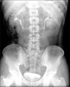
IVP results.
FEATURED BOOK: General Urology by Smith and Tanagho
When x-rays are shot at the body and a special film is placed behind the body, the x-rays
cast a shadow on the film, which produces a picture of inside the body.
Although x-rays show bones very well, any two organs in the body with similar thickness
and atomic number will be almost impossible to distinguish via x-ray.
The atomic number is the number of protons (positively charged particles) in the nucleus
(center) of an atom. An atom is the smallest part of a substance that can exist and still
possess all of the properties that are characteristic of the substance.
"Where Medical Information is Easy to Understand"™
To better distinguish between organs, a liquid substance called
contrast is used that x-rays cannot pass through. Contrast helps to
form an artificial distinction between organs in the body so that the
doctor can tell them apart. Contrast can provide this distinction
because it has a very high atomic number, unlike the lower atomic
number of the organs that it helps distinguish between.
In the case of an intravenous pyelogram, after injecting contrast into
a vein in the arm, it circulates throughout the blood and passes
through the kidneys and the rest of the urinary tract (see above).
With the contrast substance passing through these areas, the x-
rays will not be able to pass through them. it.
This will allow these areas to show up clearly on the x-ray because they will show up in contrast to areas
in the body where the x-rays easily pass through. More specifically, when x-rays hit organs that iodine has
entered, these areas will appear white on the x-ray, which serves to highlight it.
WHAT IS THE URINARY TRACT?
The urinary tract is the part of the body that deals with the formation and excretion of urine. To excrete
means to release from the body as waste. The urinary system is made up of four parts: the bladder,
urethra, kidneys, and ureters. The bladder is a stretchable structure in the body that holds urine. The
urethra is a tube shaped structure in the body that drains urine from the bladder. The kidneys are two
organs located on each side of the spine, behind the stomach. The kidneys filter (remove) wastes from
the blood. The ureters are two tube shaped structures that connect to the kidneys and carry urine to the
bladder. One ureter is attached to each kidney.
WHAT IS THE CONTRAST MADE OF?
The contrast used in an IVP is usually made of iodine (a non-metallic element) or shellfish. Yes, that's
right, shellfish. The reason for this is that seafood is the best source of iodine in the diet. Iodine is a bluish
black solid substance. Although most solids turn into a liquid and then into a gas after it is heated, iodine
does not. Rather, iodine turns directly into a purple vapor after it is heated. Because iodine does not turn
into a liquid, it needs to be combined with liquid substances so that t can be injected into the body.
WHY IS AN INTRAVENOUS PYELOGRAM PERFORMED?
An IVP is the most commonly performed test to assess if there are any problems with the urinary tract
(see above). Such problems may include injuries to the urinary tract, reoccurring urinary tract infections,
reoccurring kidney infections, kidney stones, and tumors (tissues that grow more rapidly than normal).
Cysts can also be seen, which are abnormal swellings or lumps that are filled with fluid or some solid
material. Young people with high blood pressure may be asked to get an IVP to determine if kidney
disease is a cause of the problem.
Other problems that may lead for an IVP to be done includes blood in the urine, frequent peeing, and pain
in the side or lower back. An IVP is also done if the doctor suspects that the patient may have an enlarged
prostate. The prostrate is a structure about the size of a chestnut that is below the bladder and in front of
the rear end. The prostate produces a fluid that is part of semen. Semen is a fluid that is discharged from
a male's penis in order to reproduce with a female.
HOW SHOULD I PREPARE FOR AN INTRAVENOUS PYELOGRAM?
The day before the IVP, the doctor will ask the patient to prepare for the procedure by eating less.
Depending on the patient's circumstances, the doctor will ask the patient not to eat any food for several
hours before the exam. In most cases, the patient will be asked not to eat anything after dinner or on the
night before the IVP. The patient will likely be allowed to drink clear liquids (liquids that the patient can see
through, such as water or seltzer).
Some doctors do not want the patient to drink anything for 8 to 12 hours before the IVP. Not eating or
drinking before the exam helps improve the quality of the x-ray pictures. The patient should wear
comfortable clothing since he/she will be asked to lie down on an examination table. The patient should
remove jewelry, eyeglasses, and other metal objects because they may interfere with the quality of the x-
ray pictures.
The doctor may also ask that the patient take a medication known as a laxative, such as bisacodyl. This
medication will help the patient poop. The patient may be asked to take this type of medication by mouth
and/or as a suppository. A suppository is a small, medicated mass that is shaped to be readily inserted
into another bodily opening besides the mouth.
The suppository will help the patient poop in about 15 minutes to an hour, whereas taking the medication
by mouth usually takes many hours before the patient poops. The patient may also be asked to give
him/herself an enema before the IVP. An enema is a procedure in which fluid is passed through the rear
end through a tube that is inserted in it. The enema (and the suppository) helps release poop and air from
the body.
WHAT SHOULD I BE SURE TO TELL MY DOCTOR BEFORE HAVING AN INTRAVENOUS
PYELOGRAM PERFORMED?
The patient should tell the doctor if he/she is pregnant because the x-rays can potentially harm the
developing baby. The patient should tell the doctor if he/she is taking any medications or has any
illnesses. The patient should also be sure to tell the doctor if he/she has any allergies, especially to iodine
and seafood, which is present in many types of contrast (see above).
WHAT HAPPENS DURING AN INTRAVENOUS PYELOGRAM?
After the patient checks in, he/she will be asked to take off the his/her pants or shorts and to put a hospital
gown on. The patient should be allowed to keep his/her shirt and underwear on. Before the procedure, the
patient may be asked to go to the bathroom and pee so that the strength of the contrast material is not
decreased after it mixes with the urine. Once the patient get to the examination room, he/she will be asked
to lie down on an examination table. The table should have a thin mattress on it and a pillow so that it is
comfortable for the patient. An x-ray picture will then be taken to make sure that the patient's belly is
properly prepared for the procedure. The x-ray machine is above the patient and the x-ray film is beneath
the patient.
Next, the radiologist will inject the contrast material (see above) into a vein in the arm. This may cause a
minor sting. All of the medication may be injected at once or it will slowly flow into the patient's vein
through a special hook-up. An x-ray picture will be taken immediately after the contrast is injected. In
general, x-ray pictures will also be taken 5, 10, 30, and 45 minutes after the contrast is injected. The
pictures are taken at different times to track the flow of the contrast material from the kidney to the
bladder. If the kidneys are working properly, they will begin to filter (remove) the contrast from the blood.
The contrast material may cause a warm and flushed feeling throughout the patient's body. Some patients
also notice a slightly metallic, salty taste in their mouths. These are normal reactions to the contrast and
only last for a minute or two. The patient should tell the person who is performing the IVP if he/she feels
itchy, has shortness of breath, or feels uncomfortable in any other way, because this may mean that the
patient is allergic to the contrast material. The patient should be observed by the person who is performing
the IVP to detect breathing difficulties. If breathing difficulty occurs, medication and machinery should be
readily available to help the patient breathe better.
As the contrast travels through the patient's blood, he/she may be asked to turn on his/her right and left
side for pictures to be taken while lying in these positions. When some of the x-ray pictures are taken, the
patient will be asked to hold his/her breath, but only for a few seconds. Between the 5 and 10 minute x-
rays, some pressure may be applied to the patient's belly with a tight bandage to improve the quality of the
pictures of the central cavities (openings) of the kidneys.
Toward the end of the examination, when the bladder has been filled with contrast, the patient will likely be
asked to go to the bathroom and pee. After the patient goes to the bathroom, another x-ray picture will be
taken to see whether all of the urine has left the bladder. After the x-ray pictures are taken, the patient will
be asked to wait while the pictures are reviewed to make sure they are ok. If more x-rays are needed,
they will be taken at this time.
HOW LONG DOES AN INTRAVENOUS PYELOGRAM TAKE?
An IVP usually takes about an hour. The doctor will be able to tell the patient how long his/her particular
exam will take.
WHAT HAPPENS AFTER AN INTRAVENOUS PYELOGRAM?
After an IVP, the patient will be able to go home and return to normal activities, unless told otherwise by
the doctor. The urine and feces (poop) do not usually change colors because of the contrast material (see
above). The contrast material will leave the body when the patient pees. The patient is usually encouraged
to drink more fluids to allow the contrast material to leave the body quicker and to make the contrast
material less poisonous to the kidneys. Peeing should not be painful after being injected with contrast. If
peeing is painful, or pee and/or poop have changed colors, the patient should tell the doctor right away
because it may indicate some other type of problem.
IS AN INTRAVENOUS PYELOGRAM SAFE?
Generally, an IVP is a very safe procedure. However, there are some exceptions. The procedure should
not be used with people who are very sensitive or allergic to iodine or shellfish, the main ingredient in
contrast materials (see above for more details about contrast material). People who are allergic to these
materials can have such a severe allergic reaction that it causes death. A possible way to have an IVP
done if a patient has allergies is to take anti-allergy medications before the procedure, such as
diphenhydramine hydrochloride (Benadryl). Brief hypersensitivity reactions, such as itching and nausea
are not serious and should not prevent the use of future IVPs.
Another risk of an IVP is that infections of the urinary tract can be made worse by the procedure. Patients
with serious kidney problems should not get an IVP because the kidneys will not be able to remove the
contrast material properly. Also, contrast material can be poisonous to the kidneys and cause kidney
function to worsen. Pregnant patients should avoid an IVP, especially during the first three months of
pregnancy because the x-rays may harm the developing baby. The only exception is when there is a major
risk in missing the correct diagnosis by not doing the procedure.
WHO INTERPRETS THE INTRAVENOUS PYELOGRAM?
A type of doctor known as a radiologist, that has experience in interpreting IVPs, will analyze the results
and interpret them. He or she will then write a report about the results, sign it, and send it to the patient's
doctor. The radiologist may also discuss the results with the patient's doctor. This process can be
speeded up if the place that you get the IVP done can send the results and images to your doctor over the
Internet.
WHAT CAN A RADIOLOGIST SEE ON AN INTRAVENOUS PYELOGRAM?
The pictures obtained by an IVP allow the radiologist to see the shape, size, and position of the kidneys,
the ureters, and the bladder, as well as whether there are any obvious blockages of the ureters. The
radiologist can also see the renal pelvis, which is a funnel-shaped opening that drains urine from the
kidney to the ureters. The renal calyces can also be seen, which are narrow tubes that carry urine from
the kidneys to the renal pelvis. Any blockages in these areas will cause the appearance of one or more of
the urinary tract structures to appear abnormal on the x-ray pictures.
The radiologist can also tell how well the kidneys are functioning by looking at the x-ray pictures taken
throughout the exam. If the kidneys are working properly, they will remove contrast material from the blood
and pass it into the urine, which goes to the bladder. As was mentioned earlier, the x-ray picture taken
after the patient goes to the bathroom, allows the radiologist to see whether all of the urine has left the
bladder. The radiologist can also see if the bladder is not as tense as it should be. See the beginning of
this entry for a description of the terms mentioned in this paragraph.
HOW WILL I GET THE RESULTS OF THE INTRAVENOUS PYELOGRAM?
The results of the intravenous pyelogram will be provided to the patient by the doctor that referred the
patient to get the test.
WHAT ARE THE ADVANTAGES OF AN INTRAVENOUS PYELOGRAM?
• An IVP is cheaper than other imaging procedures, such as such as magnetic resonance imaging and
computerized axial tomography.
• An IVP is a fast procedure, generally taking about an hour.
• An IVP is generally a safe procedure and complications are rare.
• An IVP requires one injection, and other than that, there is no pain or invasion of the body in another
way.
• The IVP provides your doctor with detailed information about the functioning of the urinary tract. This
information can help the doctor diagnose and treat problems in the urinary tract.
• The information obtained with an IVP can help the patient avoid surgery.
WHAT ARE SOME PROBLEMS WITH AN INTRAVENOUS PYELOGRAM?
• Some people are very sensitive or allergic to the contrast material used in an IVP, which can prevent it
from being used. However, adverse reactions to modern forms of contrast are very rare.
• In most cases, pregnant women should not have an IVP.
• Although the pictures from an IVP are clear, more clear and detailed pictures can be obtained to detect
early diseases with advanced imaging techniques such as magnetic resonance imaging and computerized
axial tomography.
• The radiation that you are exposed to during an IVP is equal to the amount of radiation that you are
exposed to in you normal environment over a period of two years. This risk can be decreased by having
high-speed films taken. These films reduce the amount of radiation that is needed for a good image to
result. Also, modern x-ray machines can decrease the amount of radiation that is spread to body parts not
being examined.
CAN I GET AN INTRAVENOUS PYELOGRAM IF I AM A BREASTFEEDING MOM?
Yes. However, the doctor or nurse will ask the mom to pump the breast milk out. The reason for this is that
iodine readily gains access to breast milk. If an infant gets too much iodine, it can lead to hypothyroidism.
Hypothyroidism is a condition in which the thyroid gland is underactive. The thyroid gland is a butterfly-
shaped organ located in front of the neck that produces hormones. Hormones are natural chemicals that
affect virtually every cell in the body and many functions such as disease fighting, heart rate, energy level,
and skin condition.
Although the breast milk should be pumped out for an IVP, the mother is not required to stop breastfeeding.
It is generally recommended by doctors that you do not breast feed for 24 hours after the procedure.
However, always speak to your doctor to check what is right timetable for your particular case.
WHAT ELSE IS AN INTRAVENOUS PYELOGRAM CALLED?
An intravenous pyelogram is also known as intravenous urography, intravenous urogram, descending
urography, and excretory urography. Intravenous pyelogram is also known as an intravenous pyelography,
which is an older term for the procedure.
WHAT IS THE ORIGIN OF THE TERM, INTRAVENOUS PYELOGRAM?
Intravenous (inside a vein) comes from the Latin word "intra" meaning "within" and the Latin word "vena"
meaning "vein." Pyelogram comes from the Greek word "pyelo" meaning "pelvis," and the Latin word
"gramma" meaning "mark." Put the words together and you get "within (a) vein, mark (the) pelvis." The
pelvis is a massive bone made of hip bones on each side and the front, while the back part is made of the
sacrum and the coccyx. The sacrum (pronounced say-crum) is a large triangle shaped bone in the lower
part of the spine. The coccyx is below the sacrum.