MedFriendly®


Template
Mastocytosis is an uncommon condition in which there
is an abnormal increase in mast cells in various organs,
most commonly the skin. The condition can be a long-
term one or it can occur suddenly.
Mast cells are types of cells that make up connective
tissue, such as tendons. Tendons are groups of fibers
that attach muscles to a bone. Mast cells are also
located in organs such as the lungs, stomach, and
intestines.
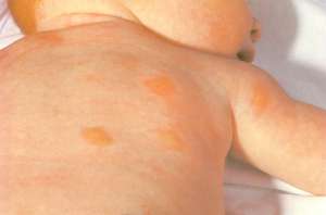
A baby with mastocytosis.
FEATURED BOOK: A Humorous Guide to Understanding Mast Cell Disorders
The lungs are two organs in the body that help people breathe. The intestine is a tube
shaped structure that is part of the digestive tract. There are more mast cells in the lungs,
intestines, and skin than in any other part of the body.
Mast cells also help to protect the body (especially the skin, stomach, and intestines)
against certain infections and diseases. Mast cells do this by releasing chemicals such
as histamine. These chemicals help attract other important aspects of the immune system
to areas of the body where they are needed. So picture mast cells as an alarm that
signals for help and which is important for survival. Histamine plays a key role in allergic
reactions, which is why mastocytosis is associated with symptoms such as flushing,
itching, swelling, and rapid heartbeat. There are certain situations that can cause a
release of histamine from mast cells and lead to symptoms of mastocytosis, such as
stress, cold, heat, insect bites, and certain medications. The triggers for histamine are
different for each person.
"Where Medical Information is Easy to Understand"™
WHAT ELSE DO MAST CELLS DO?
Mast cells also appear to play an important role in healing because
they have been shown to gather around wounds. Mast cells also
appear to play a role in the growth of blood vessels. It appears that
people would not be able to survive without enough mast cells.
WHAT CAUSES MASTOCYTOSIS?
by changes in the bodys immune system. These changes result
from the release of chemicals produced by too many mast cells.
The exact cause of the increase in mast cells is not known. However, at least in some cases, the
increase in mast cells is thought to be due to a response to an unknown stimulus.
A substance produced by the body known as mast cell growth factor has been shown to increase the
amount of mast cells that are produced. Mast cell growth factor also increases the amount of melanocyte
and melanin pigment (colored substance) that is produced, which explains the colored areas that are
found in abnormal tissue areas in mastocytosis that affects the skin. A melanocyte is a cell that is able to
produce melanin. Melanin is a yellow, brown, or black colored substance that gives coloring to the hair,
skin, and the iris (colored part of the eye).
The increase in melanin is found in the lowest layer of the epidermis. The epidermis is the top layers of
the skin. Substances that eat melanin, called melanophages, have been found in the upper part of the
dermis (the layer of skin below the epidermis) and contribute to the increased coloring of the skin found in
skin mastocytosis.
Other substances that lead to mastocytosis symptoms include histamine (see above), heparin (a blood
thinner), prostaglandins, acid hydrolases, and neutral proteases. Prostaglandins are types of fatty acids
made naturally by the body that act like hormones. Hormones are natural chemicals produced by the body
and released into the blood that have a specific effect on tissues in the body.
Acid hydrolases are types of enzymes. An enzyme is a type of protein that helps produce chemical
reactions in the body. A neutral protease is a type of enzyme that helps breakdown certain parts that
makeup a protein. The term pH stands for potential hydrogen and is used to determine how acidic a
liquid is, such as the blood. A pH below 7 is acidic, above 7 is alkaline (opposite of acidic), and 7 is
neutral. A neutral protease is given this name because it is only active at a neutral pH.
Symptoms of mastocytosis can develop in the bones, heart, lungs, brain, spine, stomach, and intestines, if
mast cells enter these areas. The release of the substances described in this section can also cause
symptoms in these areas as well. Severe allergic reactions can also result causing fainting, abnormally
low blood pressure, and (if not treated promptly) death. Severe allergic reactions are caused by the
sudden and severe release of tiny substances (known as granules) from many mast cells. This has been
known to happen in adults with mastocytosis after an insect sting.
CAN MASTOCYTOSIS BE GENETIC?
It is possible that there are some rare cases of urticaria pigmentosa (the most common type of skin
mastocytosis) that are genetic, meaning that they are passed on from generation to generation through
genes. Genes are units of material contained in a person's cells that contain coded instructions as for
how certain bodily characteristics (such as eye color) will develop.
WHAT ARE OTHER SIGNS & SYMPTOMS OF MASTOCYTOSIS?
The symptoms of mastocytosis vary between individuals. The symptoms differ depending on where the
high levels of mast cells are. When the skin is involved, the most common reported symptom is itchiness.
The coloring of the skin caused by mastocytosis tends to be yellow-tan to reddish-brown. The affected
areas can be small, flat, and level with the skin. The affected areas can also be small, raised, and less
than one centimeter in width. Small, rounded masses can also occur. It should be noted that the affected
areas of the skin can be less than a millimeter (one thousandth of a meter) to several centimeters in width.
Rarely, mastocytosis can cause the formation of destructive lesions (abnormal areas in body tissue) that
have thick, hardened margins. Hardened areas of the skin can occur throughout the body.
The number of noticeably affected areas on the skin varies from 1 to 1000. If both areas of the body are
affected, both sides tend to be affected equally. The trunk tends to be affected more than the arms and
legs. The trunk refers to all the parts of the body, except the head, arms, and legs. The face, scalp, soles
of the feet, and palms tend to be spared.
Other common symptoms of skin mastocytosis include redness, a lump under the skin, and/or rashes with
a freckle-like appearance. The lump under the skin can be caused by the mast cells building up in one
particular area. In infancy, the bumps on the skin can become filled with fluid.
Rubbing a rash or affected area of the skin can cause the skin to become more red, itchy, and swollen.
When this happens it is known as the Darier sign and is caused by the release of lysosomes from a cell
after physical stimulation. Lysosomes are particles that contain enzymes which digest cells inside the
body. The Darier sign does not usually occur in a type of mastocytosis known as paucicellular
mastocytosis. Paucicellular mastocytosis is an eruption of widened blood vessels in the skin associated
with red and swollen bumpy masses. Eating or drinking certain substances can cause a release of
lysosomes, which can lead to flushing of the skin. A sudden, widespread loss of lysosomes can lead to
life-threatening episodes of shock. In about half of affected patients, stroking large areas of uninvolved
skin will produce a red mark(s).
Mast cells in the stomach can cause diarrhea and stomach pain, but this is rare. Other symptoms of
mastocytosis include a general feeling of sickness, pain in the muscles and bones, bone fractures,
abnormal thickness of bone tissue throughout the body, shock, headache, dizziness, nausea, vomiting,
belly cramps, lack/loss of appetite, weight loss, and stomach ulcers. An ulcer is an open sore on the skin
or on a mucous membrane. A mucous membrane is one of four major types of thin sheets of tissue that
line or cover various parts of the body, such as the mouth and passages for breathing.
In some people, mastocytosis can cause symptoms that are like a severe allergic reaction. Specifically,
the blood pressure can drop, causing the person to faint. Difficulty breathing, runny nose, wheezing, death
can occur if the person is not immediately treated. This type of reaction, however, is rare. In cases where
the heart is affected, rapid heart beat, episodes of low blood pressure, and fainting can result. The fainting
results from widening of the blood vessels.
Enlarged glands can occur in mastocytosis. A gland is an organ in the body made of special cells that form
and release materials such as fluid. The liver and spleen can become enlarged in mastocytosis in about
40 and 50% of cases, respectively. This is because of increased amounts of mast cells that are present
in these areas.
The liver is the largest organ in the body and is responsible for filtering (removing) harmful chemical
substances, producing important chemicals for the body, and other important functions. The spleen is an
organ near the stomach that helps fight infection and removes and destroys worn-out red blood cells. Red
blood cells help carry oxygen to the blood.
Anemia can occur in mastocytosis. Anemia is a condition in which there is an abnormally low amount of
hemoglobin in the blood. Hemoglobin is substance present in red blood cells that help carry oxygen to cells
in the body. Leukocytosis can also occur, which is an abnormal increase in the number of white blood
cells. An increase in a type of white blood cell, known as eosinophils, can also occur in mastocytosis.
Thrombocytopenia can occur in mastocytosis. Thrombocytopenia is a reduction of the number of platelets
in the blood. Platelets are cells that help stop bleeding by changing the blood from a liquid to solid form.
Mastocytosis can also cause either a decrease or increase in the number of white blood cells. White
blood cells help protect the body against diseases and fight infections. Psychological symptoms can also
occur such as irritability.
ARE THERE DIFFERENT TYPES OF MASTOCYTOSIS?
Yes. Mastocytosis can occur in and affect various organs throughout the body. When this happens, it is
referred to as systemic mastocytosis. Systemic mastocytosis is the more severe form of this disease.
Mastocytosis can spread to the bones, the lining of the stomach, intestine, the liver, spleen, and bone
marrow. Bone marrow is a type of tissue that fills the inside of bones.
Mastocytosis can also occur more locally, in the skin, appearing as a spotted rash. This is often known as
cutaneous mastocytosis (CM) or skin mastocytosis. The most common type of skin mastocytosis is
urticaria pigmentosa, which was first reported in the scientific literature in 1933. It is characterized by
many small reddish-brown bumpy areas on the skin, ranging from just a few to thousands. These skin
abnormalities, when seen in children, can sometimes be misinterpreted as signs of physical abuse.
Other types of skin mastocytosis are solitary mastocytoma, diffuse erythrodermic mastocytosis, and
paucicellular mastocytosis (also called telangiectasia macularis eruptiva perstans or TMEP). A solitary
mastocytoma is a well-defined buildup of mast cells in a specific part of the body that resembles a bumpy
mass. Diffuse erythrodermic mastocytosis is a condition in which there are flat or slightly elevated areas
on the skin that itch when touched. Paucicellular mastocytosis is an eruption of widened blood vessels in
the skin associated with red and swollen bumpy masses. In paucicellular mastocytosis, mast cells usually
are not present in enough numbers for touching of the skin to cause itchiness
CAN MASTOCYTOSIS AFFECT ANY PART OF THE BODY?
Yes, mastocytosis can affect any part of the body but it most commonly affects the trunk. The trunk refers
to all the parts of the body, except the head, arms, and legs.
WHEN DOES MASTOCYTOSIS USUALLY OCCUR?
Mastocytosis usually begins in the first year of life and disappears by adolescence.
IS MASTOCYTOSIS A RISK FACTOR FOR CANCER OR OTHER ILLNESSES?
In some cases, mastocytosis can be a risk factor for cancer. Specifically, mastocytosis may occur before
mast cell leukemia. Leukemia is a type of cancer in the blood in which bone marrow (a tissue that fills the
openings of bones) is replaced by early forms of white blood cells. In mast cell leukemia, abnormal
collections of mast cells from connective tissue are present in circulating blood. Mastocytosis is
considered to be a risk factor for possibly developing a serious blood disorder.
WHO GETS MASTOCYTOSIS?
Mastocytosis usually occurs in white people. In people with many freckles or other naturally colored areas
of the skin, the increased skin coloring caused by most types of mastocytosis will not be very noticeable.
Mastocytosis can happen to both children and adults and it can occur at any age. However, about 75% of
cases occur in infancy or early childhood. The amount of people that develop mastocytosis increases
again in people between the ages of 30 and 49. Males and females tend to be equally affected.
Mastocytosis in childhood is usually mild and the children tend to outgrow it. However, mastocytosis is
usually more serious in adults. Cutaneous mastocytosis mostly affects children.
HOW MANY PEOPLE HAVE MASTOCYTOSIS?
Mastocytosis is considered an orphan disease, meaning that it affects fewer than 200,000 people in the
United States. It is possible that mastocytosis may be misdiagnosed as some other kind of disorder and
that there really are more than 200,000 cases. In the U.S., it is estimated that about 0.1 to 0.8% of new
patients that visit a skin doctor have some form of mastocytosis.
HOW IS MASTOCYTOSIS DIAGNOSED?
The skin version of mastocytosis is diagnosed after a physician performs a physical examination of the
skin and sees the characteristic color changes. The presence of the Darier sign indicates the presence of
mastocytosis. The Darier sign is when rubbing an affected area on the skin can cause the skin to become
more red, itchy, and swollen. The diagnosis can be difficult because the symptoms can be like those of
many other medical problems. A procedure known as a biopsy needs to be performed to confirm the
diagnosis. A biopsy is a procedure in which a skin sample is taken from an affected organ or body part
and sent for analysis in the laboratory.
As an example of a biopsy, samples of bone marrow can be taken to be analyzed to confirm the diagnosis.
Bone marrow is a tissue that fills the openings inside bones. Bone marrow biopsies are more common in
adults with mastocytosis. Your doctor may consult with a hematologist (a doctor that specializes in blood
and blood disorders) for the bone marrow biopsy, especially if the blood count results are abnormal or
there are abnormal findings from radiologic techniques. Radiographic (or radiologic) examinations are
techniques (such as x-rays) that use radiation (a type of energy) to generate pictures of the inside of the
body.
A bone marrow aspirate can also be performed, in which fluid is taken out of the bone marrow. The bone
marrow biopsy and aspirate are done to look for other diseases that may be present along with
mastocytosis, such as blood disorders. They are often done in patients that have urticaria pigmentosa
(the most common type of skin mastocytosis) that also have abnormal blood test results, an enlarged
liver, enlarged spleen, or an enlarged lymph node(s). Lymph nodes are small egg shaped structures in the
body that help fight against infection.
To do the biopsy, the doctor will inject anesthesia (pain blocking medication) next to, not into, the
abnormal tissue area. Not injecting the needle into the abnormal tissue helps prevent it from releasing
lysosomes, which would make the laboratory analysis difficult. See the earlier section for a discussion of
lysosomes.
Under the microscope, increased numbers of mast cells will be seen in the tissue samples of
mastocytosis patients, especially near the blood vessels of the outer projections of the dermis. The
dermis is the layer of skin below the epidermis. The epidermis is the top layers of the skin. Certain stains
are used on the tissue samples to help see the mast cells better. Some common stains used to detect
mast cells are Giemsa, Leder, and toluidine blue stains. The Leder stain is able to detect mast cells that
are not intact.
Mast cells tend to be spindle shaped in cases of urticaria pigmentosa which presents with abnormal
tissues areas that are small, flat, and level with the skin or in cases in which the abnormal tissue is small
(less than one centimeter in width), solid, and raised. Mast cells tend to be cube-shaped in cases of
urticaria pigmentosa with affected skin areas that are small, firm, and knotty. In this type of mastocytosis,
the mast cells are observed in big groups and can extend throughout the dermis (see last paragraph) and
into layers beneath the skin.
If the affected skin were traumatized when the sample was taken, a buildup of excessive fluid and the
presence of eosinophils (a type of white blood cell) may be found in the sample.
Another way to diagnose mastocytosis is to have an analysis performed of the blood or urine to look for
high levels of proteins or high levels of chemicals (such as histamine) that are produced by mast cells.
This is likely to be done if the patient has a rash, but does not have other symptoms. Patients with
extensive areas of affected skin may have histamine levels in the urine that are 2 to 3 times the normal
level.
In addition to testing for histamine levels, there are three different substances that can be tested for with
blood and/or urine tests to help diagnose mastocytosis. The first substance that can be tested for is the
level of tryptase in the blood, which elevated in patients with mastocytosis. Tryptase is a type of enzyme
that splits the molecules of certain proteins. An enzyme is a type of protein that helps produce chemical
reactions in the body. Molecules are the smallest naturally occurring particles of a substance.
Two other substances, known as N-methylhistamine (NMH) and N-methylimidazoleacetic acid can be
tested in the urine. These two substances are the two major metabolites of histamine found in the urine.
Metabolites are substances produced by metabolism. The levels of NMH are associated with the extent of
abnormal tissue areas in mastocytosis.
Another substance that can be tested for (although the test is not widely available) are metabolites of
urinary prostaglandin D2. Metabolism is the chemical actions in cells that release energy from nutrients or
use energy to create other substances. Urinary prostaglandin D2 is a type of fatty acid found in the urine
that is naturally made by the body. Fatty acids are types of chemicals in the body that are necessary for
many bodily functions. Metabolites are produced when urinary prostaglandin D2 does its job. Even when
patients with mastocytosis are not having symptoms, metabolites of urinary prostaglandin D2 can be
found up to 150 times the normal level.
The doctor may want the patient to have a bone scan performed in adults or in children with an abnormal
complete blood count (CBC). A bone scan is a special technique used to produce pictures of bones. The
CBC test is performed to see if an infection is present. If an infection is present, certain types of white
blood cells will be abnormally increased in number.
If the patient has complaints about symptoms relating to the bones, a bone scan is usually also performed.
The bone scan can detect abnormal areas of bone that can be due to multiple causes such as
osteoporosis or osteosclerosis. Osteoporosis is an abnormal loss of bone thickness and a wearing away
of bone tissue. Osteosclerosis is an abnormal hardening or thickening of bone, which sometimes can
resemble ivory in appearance.
If symptoms occur in the digestive tract, additional tests such as x-rays of the affected area may be
performed to determine if there are other problems contributing to the symptoms.
HOW IS MASTOCYTOSIS TREATED?
Treatment of mastocytosis is difficult and there is no cure. The goal is to treat the patients specific
symptoms and to stay away from situations or substances (such as alcohol) that trigger the symptoms to
begin with.
Antihistamines have also been shown to be of some help, especially in decreasing itchiness, redness, and
digestive symptoms (such as belly pain and diarrhea). Antihistamines are substances that block the
effects of histamine and are often used to treat allergies. Histamine is a natural substance in the body that
is released during allergic reactions and leads to many allergic symptoms. Histamine is released by mast
cells.
Histamine exerts its effect by interacting with three different types of histamine receptors in the body,
known as H1, H2, and H3. Antihistamines block these receptors, thus preventing histamine from
interacting with them. This, in turn, blocks the effect of histamine. Antihistamines that block the H1 and H2
receptors are most often used to treat mastocytosis. In fact, many patients with skin mastocytosis are
started on H1 antihistamines, which are given by mouth.
H2 antihistamines are sometimes added to H1 antihistamines to control itching, stomach distress, and
types of skin eruptions known as wheals. Because mastocytosis can cause severe allergy symptoms, it is
a good idea to keep an emergency kit so medication can be taken in cases of a severe reaction.
An example of an anti-allergy drug that has been found to be helpful in relieving certain symptoms of
mastocytosis is disodium cromoglycate (aka Cromolyn sodium or Gastrocrom). The medication works by
preventing the release of particles from mast cells that lead to symptoms. When taken by mouth, this
medication has been shown to decrease symptoms of skin mastocytosis such as itching, flushing, and
types of skin eruptions known as wheals. Disodium cromoglycate has also been found to be helpful in
reducing signs of systemic mastocytosis, such as diarrhea, vomiting, belly pain, bone pain, and thinking
disorders. It is often given with H1 antihistamines (seen above) but is sometimes given alone at the
beginning of treatment for patients with skin mastocytosis.
For patients whose bodies resist the effects of H1 and H2 antihistamines, some doctors have found it
helpful to cautiously administer small doses of aspirin. Aspirin can help decrease the production of
prostaglandins. Prostaglandins are types of fatty acids made naturally by the body that act like hormones.
Prostaglandins have been known to trigger mastocytosis, but so has aspirin, which is why it needs to be
given cautiously. Both prostaglandins and histamine are types of mast cell mediator, which are types of
chemicals that can cause allergic reactions.
Skin lesions that are present in a limited area of the body can be treated with a class 1 (mildly potent)
corticosteroid that can be rubbed on the affected area. Corticosteroids are a group of drugs that act
similarly to a natural chemical in the body known as corticosteroid hormone. Corticosteroid hormones
control the body's use of nutrients and the amount of water and salts in the urine. Corticosteroids may or
may not be used to treat selected skin abnormalities in the beginning of treatment for skin mastocytosis.
Skin lesions can also be injected with small amounts of corticosteroids that have been diluted. This can
cause the skin lesions to go away temporarily or indefinitely. Only limited areas of the body would be
treated this way in a single session to decrease the chance of an abnormal loss of skin tissue in the area
of the injection. Limited treatments with corticosteroid injections help decrease the chances of
adrenocortical suppression. The adrenal glands are a pair of glands that play an important role in
metabolism and help the body respond to physical and emotional stress by releasing certain hormones.
Glands are organs in the body made of special cells that form and release materials such as fluid.
Metabolism is the chemical actions in cells that release energy from nutrients or use energy to create
other substances.
Corticosteroids are only administered to treat the entire body in specific situations, such as severe skin
disease, malabsorption, and ascites. Ascites is a condition in which fluid builds up in the space between
layers that line the belly. A build up of fluid in this area will cause the belly to look swollen.
Medicines that help relieve cramping in the intestines have also been helpful in treating symptoms of
mastocytosis. Other specific medications can also be used to treat symptoms such as itching, other skin
reactions, diarrhea, poor nutrition, and low blood pressure. If mastocytosis is associated with a blood
disorder or cancer, medications will obviously be needed to treat those conditions.
A treatment known as PUVA is sometimes used to treat skin rashes and other skin problems associated
with skin mastocytosis. PUVA is especially useful in the treatment of TMEP (see above). The P in PUVA
stands for psoralen, which is a medication that contains chemicals that react with ultraviolet light, causing
darkening of the skin. This is where the UVA comes in, which stands for a type of ultraviolet light. So
PUVA is a combination of Psoralen and a type of ultraviolet light. Risks of PUVA include skin cancer,
especially if more than 200 treatments are needed. Children rarely need PUVA because it is a treatment
that is typically reserved for severe unresponsive cases.
For patients with systemic mastocytosis or extensive symptoms, a drug known as epinephrine is often
administered. Epinephrine helps relieve the body of severe allergic reactions by causing the blood vessels
to narrow. In severe allergic reactions, the blood vessels are too wide, causing the blood pressure to be
too low since it is not moving well. By causing the blood vessels to narrow, the blood can move throughout
the body better, causing blood pressure to improve. In the most severe allergic reactions, emergency
resuscitation may be needed if the patient faints or goes into a state of shock with abnormally low blood
pressure.
SHOULD PEOPLE WITH MASTOCYTOSIS FOLLOW A SPECIAL DIET?
Patients with mastocytosis are often advised by their doctors to avoid substances that lead to the release
of mast cell mediators, which are types of chemicals that can cause allergic reactions. Such substances
include alcohol, lobster, crawfish, cheese, spicy foods, and hot drinks.
SHOULD PEOPLE WITH MASTOCYTOSIS AVOID CERTAIN MEDICATIONS?
People with mastocytosis are often advised to stay away from salicylates. Salicylates are any of several
widely prescribed drugs derived from salicylic acid, an example of which is aspirin. Salicylic acid is a
substance that causes anti-fever, anti-inflammatory, and pain-relieving responses.
Other medications that patients with mastocytosis are often advised to stay away from includes codeine,
procaine, opiates, and NSAIDs (non-steroidal anti-inflammatory drugs), which are all used to treat pain.
Aspirin can trigger the release of mast cell mediators, which are types of chemicals that can cause
allergic reactions. The release of mast cell mediators can eventually lead to a heart failure. Other
medications that can trigger mastocytosis include thiamine (a type of vitamin), polymyxin b (an antibiotic),
and scopolamine (used to treat motion sickness). Three types of muscle relaxants, gallamine,
decamethonium, and D-tubocurarine can all trigger mastocytosis symptoms.
Quinine can also trigger mastocytosis symptoms. Quinine is a white, bitter tasting powder that is obtained
from the bark of the cinchona tree. Quinine is found in the clear drink, tonic. Dextran, a sugary substance,
can trigger mastocytosis symptoms. Dyes given during radiographic examinations can also trigger
mastocytosis. These dyes are substances that help highlight areas of potential abnormality on the
pictures that result from the radiographic examinations. Radiographic (or radiologic) examinations are
techniques (such as x-rays) that use radiation (a type of energy) to generate pictures of the inside of the
body.
SHOULD PEOPLE WITH MASTOCYTOSIS RESTRICT THEIR ACTIVITIES IN ANY WAY?
Patients with mastocytosis are often advised by their doctors to avoid certain physical stimuli, including
physical exertion, emotional stress, temperature extremes, poisons from bacteria, and venom from
insects that he/she is allergic to. Rubbing, scratching, bathing, and/or causing any trauma to the lesions in
skin mastocytosis should be avoided.
WHAT OTHER PRECAUTIONS CAN PEOPLE WITH MASTOCYTOSIS TAKE?
People with systemic mastocytosis or who have extensive symptoms can wear medical alert bracelets in
case a severe allergic reaction occurs that leads to shock. These individuals can also carry injectable
epinephrine (see above).
WHAT IS THE PROGNOSIS FOR MASTOCYTOSIS?
There is no cure for mastocytosis. The prognosis depends on the age that symptoms began. Prognosis is
usually excellent if the symptoms are treated and the person stays away from situations that trigger the
symptoms. For the most common type of mastocytosis, urticaria pigmentosa (UP), most cases that occur
in childhood (typically before age 2) go away spontaneously before puberty. The number of skin
abnormalities is usually reduced by 10% per year.
Patients with UP that began in adolescence or early adulthood (after age 10) are more likely to have signs
of persistent disease and are greater at risk for the condition to involve several areas of the body. Thus,
the prognosis for these patients is poorer. Mastocytosis becomes malignant (meaning that it spreads and
invade surrounding tissues) in about 7% of cases in which the condition begins before adulthood. By
contrast, mastocytosis becomes malignant in about 30% of adult cases in which the condition begins in
adulthood.
WHO FIRST DESCRIBED MASTOCYTOSIS?
Mastocytosis was first described by E. Nettleship and W.Tay in 1869. The title of the article was of Rare
forms of Urticaria" and was published in the British Medical Journal. Incidentally, tissue mast cells were
first identified by Paul Ehlrich in 1877. Systemic mastocytosis was first described by scientists in 1936. In
1949, Dr. J.M Ellis performed an autopsy on a patient with mastocytosis and proved that this condition
can spread to the internal organs.
WHAT IS THE ORIGIN OF THE TERM, MASTOCYTOSIS?
Mastocytosis comes from the German word "mast" meaning "fattening," the Greek word kytos meaning
cell, and the Greek word "osis" meaning "condition." Put the words together and you have "fattening cell
condition."