MedFriendly®


Hydronephrosis
Hydronephrosis is a condition in which specific
parts of the kidney(s) become stretched,
swollen, and widened with urine. The kidneys
are two organs located on each side of the
spine, behind the stomach. The kidneys filter
(remove) wastes from the blood.
FEATURED BOOK: Idiopathic Hydronephrosis
WHAT PARTS OF THE KIDNEYS ARE WIDENED IN HYDRONEPHROSIS?
The parts of the kidney(s) that are widened in hydronephrosis are the renal calyces and
renal pelvis. The word "renal" refers to the kidneys. The renal calyces are narrow tubes
that carry urine from the kidneys to the renal pelvis. The renal pelvis is a funnel-shaped
opening that drains urine from the renal calyces to the ureters. The ureters are two thin,
tube shaped structures that connect to the kidneys and carry urine to the bladder. One
ureter is attached to each kidney. The bladder is a stretchable structure in the body that
holds urine.
Urine travels from the kidneys to the renal calyces, then to the renal pelvis, then to the
ureters, then to the bladder, and then to the urethra where it is drained outside of the
body. The urethra is a tube shaped structure in the body that drains urine from the
bladder. The kidneys, ureters, bladder, and urethra make up the urinary tract. The
widening in hydronepohrosis is most common at the point where the renal pelvis connects
with the ureters.
"Where Medical Information is Easy to Understand"™
IS HYDRONEPHROSIS A DISEASE?
Hydronephrosis is not a disease. Hydronephrosis is a physical
consequence of a disease (see below) that causes the urine to
have difficulty draining out of the kidneys, ureters, and bladder.
WHAT IS THE DIFFERENCE BETWEEN UNILATERAL AND
BILATERAL HYDRONEPHROSIS?
Unilateral hydronephrosis is hydronephrosis that affect one kidney.
Bilateral hydronephrosis is hydronephrosis that affects both
kidneys.
WHAT ARE SIGNS AND SYMPTOMS OF HYDRONEPHROSIS?
Signs and symptoms of hydronephrosis can vary, depending on whether the condition occurs suddenly or
develops slowly over a period of time. If hydronephrosis occurs suddenly, the person may experience
severe back pain.If hydronephrosis develops slowly over a period of time, it may not cause any signs or
symptoms until the ureter is completely blocked and kidney failure results. If the kidney becomes infected,
it can become filled with pus and lead to a fever. Pus is a yellow or green creamy substance sometimes
found at the site of infections.
General signs and symptoms of hydronephrosis include pain in the sides, a mass in the belly, very high
fever, blood in the urine, nausea, vomiting, and a high number of white blood cells in the urine. White blood
cells help protect the body against diseases and fight infections. In the newborn, an infection of the
urinary tract is a sign that some type of blockage in the kidney is present that may lead to hydronephrosis.
In older children, repeated infections of the urinary tract may reflect a blockage of the kidney.
Feeling a frequent urge to pee and peeing very often are other signs and symptoms of hydronephrosis.
Painful and burning sensation while peeing is another symptom of hydronephrosis that is usually caused
by an infection of the urinary tract. It should be noted that in many people, there are no signs or symptoms
of a blockage in the urinary tract. This is especially true for children with mild hydronephrosis.
WHAT IS NEONATAL (CONGENITAL) AND ANTENATAL HYDRONEPHROSIS?
Neonatal hydropnephrosis (also known as congenital hydronephrosis) is hydronephrosis that occurs in
newborns. Antenatal hydronephrosis is hydronephrosis that is detected before the birth of the child. There
is no evidence that congenital hydronephrosis is due to anything that the parents did wrong during
pregnancy.
WHAT CAUSES HYDRONEPHROSIS?
In hydronephrosis, the kidneys become widened with urine (pee) that cannot pass by a blockage or
narrowing. The blockage or narrowing is usually in the ureter or urethra, but can also be in the bladder.
Blockages can occur at the uretovesical junction, which is where the ureter meets the bladder. Since the
urine has difficulty getting past the blockage, it backs up into the kidney and becomes trapped. This
causes areas inside the kidney to stretch. The blockages can occur suddenly or slowly over time. In
infants with this condition, the areas inside of the kidney may appear very wide because the infant's
tissues are stretchier than adults
There are many possible reasons why the structures mentioned above can become blocked or narrowed.
One reason is the formation of stones in the kidney (known as kidney stones). Another reason is the
formation of scar tissue in the ureter or urethra. A blockage where the bladder connects to the urethra can
lead to hydronephrosis. This type of blockage is known as bladder outlet obstruction.
A tumor in the kidney or other parts of the urinary tract can cause blockage of the ureter(s). Tumors are
abnormal masses of tissue that form when cells in a certain area of the body reproduce at an increased
rate. Tumors outside or in back of the peritoneum can lead to hydronephrosis. The peritoneum is a layer
of tissue that lines the belly and covers most of the organs in the belly.
As was mentioned earlier, the widening in hydronepohrosis is most common at the point where the renal
pelvis connects with the ureters (known as the uretopelvic junction). A blockage in the area where the
renal pelvis connects with the ureters can cause widening in this area to occur. A blockage in this area is
known as a uretopelvic junction obstruction. A uretopelvic junction obstruction usually develops in the first
four months that a mother is pregnant, in children born with this type of blockage.
The presence of abnormal folds in a specific part of the urethra can lead to hydronephrosis. This condition
is known as posterior urethral valves. Also, fibrosis of structures behind the peritoneum and of connective
tissue that often makes up the ureters can lead to hydronephrosis because it can cause the ureters to be
blocked. Fibrosis is an overgrowth of scar tissue or connective tissue. Connective tissue is any tissue
that surrounds and supports specialized structures in the body.
Displacement of the kidneys from its normal position can lead to hydronephrosis as can pressure from
adhesions outside of the urinary tract. Adhesions are fibrous types of tissue that join body parts that are
normally unconnected. Fibrous tissue is a type of connective tissue.
Sometimes, the kidney may contain many cysts, which can lead to hydronephrosis. A cyst is an abnormal
lump, swelling, or sac that contains fluid, a part solid material, or a gas, and is covered with a membrane.
A blood clot can also lead to a blockage of the ureters and cause hydronephrosis. A blood clot is a
collection of a mass of blood.
Pressure from abnormal arteries in the kidneys can lead to hydronephrosis. An artery is a blood vessel
that carries blood away from the heart. Aneurysms of the arteries of the kidney are one type of
abnormality that can lead to hydrnephrosis. An aneursym is a weakening of the wall of a blood vessel,
causing it to expand like a balloon, sometimes leading it to burst.
Neurogenic bladder can lead to hydronephrosis. Neurogenic bladder is dysfunction of the bladder that is
caused by damage in the nervous system (the system of nerves in the body). Neurogenic bladder is
characterized by retaining or emptying too much urine.
Another cause of hydronephrosis is an enlarged prostate gland. The prostate is a organ near the bladder
that produces a reproductive fluid. If the prostate is enlarged, the fluid that it produces can be blocked,
which would cause the urine in the bladder to be under pressure.
Cancer of the prostate gland can cause blockage that leads to hydronephrosis. Cancer is an abnormal
growth of new tissue characterized by uncontrolled growth of abnormally structured cells that have a more
primitive form. One type of cancer in the prostate that can lead to hydronephrosis is a carcinoma.
A carcinoma is any malignant cancer that arises from cells in the covering surface layer or membrane
(outer covering) that lines an organ of the body. When describing cancer, malignant means that the newly
formed tissues are made of abnormally structured and primitive-looking cells that grow uncontrollably,
spread throughout the body, and invade surrounding tissues.
Vesicouretal reflux is another possible cause of hydronephrosis. Vesicouretal reflux is a backward flow of
urine from the bladder into the ureter, when the bladder fills or empties. The urine can also flow black into
the renal pelvis. This condition is caused when the valve between the ureter and bladder does not
function. A valve is a natural structure or man-made device in a passageway, tube, vessel, or hollow
organ that allows fluid or partly fluid contents to travel in one direction, but closes to prevent the flow of
those contents in the opposite direction.
About 75% or people with vesicouretal reflux outgrow this condition during childhood. However, many
children will need to be treated with antibiotics to prevent kidney damage before outgrowing the condition.
Edema that is caused by an infection of the urinary tract can lead to hydronephrosis. Edema is an
abnormal buildup of too much watery fluid in the cells, tissues, and or openings inside the body. Pregnancy
can cause changes in the urinary tract that lead urine to be backed up, which can cause hydronephrosis.
A condition known as prune-belly syndrome is associated with hydronephrosis. In prune belly syndrome,
certain muscles in the belly are missing, the kidneys are small and abnormally developed, the testicles do
not drop down, and the ureters and bladder are usually greatly enlarged. The condition is called prune-belly
syndrome because the belly sticks out and is wrinkly like a prune.
There are several disorders of the muscles, brain, and/or spinal cord that can lead to hydronephrosis,
such as paraplegia, tabes dorsalis, spina bifida, and multiple sclerosis. Paraplegia is a loss of the ability
to move and/or feel both legs and generally, the lower trunk (stomach area and lower back). Tabes
dorsalis is damage to the spinal cord due to infection from the sexually transmitted disease, syphilis.
Spina bifida is a birth defect in which part of the bones that surround the spinal cord do not form properly,
leaving part of the spinal cord exposed. Multiple sclerosis (MS) is a condition in which multiple areas of
abnormal patches (known as plaques) develop in the brain and/or spinal cord (depending on the stage of
the illness).
Sometimes, in adults and children, the ureters can become narrowed at the area of the renal pelvis with
no apparent cause. In some children with hydronephrosis, there is no blockage that is apparent. Some of
these cases may be due to abnormal smooth muscle in the renal pelvis or ureter, which causes these
areas to widen.
A rare cause of hydronephrosis can occur when there are two ureters collecting urine from a single
kidney. In 99% of people, there is one ureter that collects urine from each kidney. In the 1% of people that
have 2 ureters for one kidney, a uterocele may be present which causes hydronephrosis. A uterocele is a
balloon-like blockage at the end of one of the ureters.
HOW MANY PEOPLE HAVE HYDRONEPHROSIS?
It is estimated that one in 100 people have hydronephrosis affecting one kidney. Less people have
hydronephrosis affecting both kidneys. In approximately 2% of all ultrasounds performed before the child
is born, hydronephrosis is detected. The cases detected in this manner are usually males.
HOW IS HYDRONEPHROSIS DIAGNOSED?
Hydronephrosis is diagnosed with an ultrasound of the kidneys or belly, especially in unborn children. An
ultrasound is a procedure that uses types of sound waves to produce images of the body. The ultrasound
used to diagnose hydronephrosis will produce a picture of the kidneys and the ureters. An ultrasound can
detect hydronephrosis in an unborn child as early as the first three months that the mother is pregnant. It
will be able to show blockages that are causing hydronephrosis as well as many of the other causes
described above, such as uretopelvic junction obstruction, posterior urethral valves, and prune belly
syndrome.
Another technique used to diagnose hydronephrosis is an intravenous pyelogram. An intravenous
pyelogram (commonly abbreviated as IVP) is a technique in which x-rays are used to take pictures of the
urinary tract, after a liquid substance called contrast is injected into a vein (a blood vessel that caries
blood to the heart). X-rays are a type of radiation, which is a form of energy. To better distinguish
between organs, a liquid substance called contrast is used that x-rays cannot pass through. Contrast
helps to form an artificial distinction between organs in the body so that the doctor can tell them apart.
Other techniques that can be used to diagnose hydronephrosis are retrograde pyelography and
cystoscopy. A retrograde pyelography is another visualization technique that uses x-rays to examine
structures in the kidneys after a contrast material has been injected into the ureters and renal calyces. A
cystoscopy is a direct examination of the urinary tract by inserting a viewing tube into the ureter.
A renal (kidney) scan is also used to diagnose hydronephrosis in some cases. A renal scan (also known
as a flow and function scan) is a technique in which detailed pictures are produced after a radioactive
substance is injected into the body, which provides information about kidney functioning. A radioactive
substance is a substance that sends out radiation (a type of energy). The kidney filters this radioactive
substance, which is detected by the scanning procedure.
The renal scan will be able to detect differences in functioning between the right and left kidney. It can
estimate the size of a blockage that is present and how well the urine is draining out of the kidney. A small
tube is inserted in the bladder to keep it empty. Renal scans are usually not performed until one month
after birth for the most accurate results.
A CT scan of the kidneys or belly can be used to diagnose hydronephrosis. CT (computerized
tomography) scanning is an advanced imaging technique that uses x-rays and computer technology to
produce more clear and detailed pictures than a traditional x-ray. An MRI (Magnetic Resonance Imaging)
scan can also be used to diagnose hydronephrosis. MRI scans produce extremely detailed pictures of the
inside of the body by using very powerful magnets and computer technology. MRI scans are more
detailed, but more expensive than CT scans.
A cystourethogram can be used to help determine if the cause of hydronephrosis is vesicourethral reflux.
A cystourethogram is an examination of the urethra and bladder after a contrast substance is injected
inside the body. Remember that contrast is a liquid substance that helps to form an artificial distinction
between organs in the body so that the doctor can tell them apart. The contrast is passed into the child's
bladder through a small tube inserted in the child's urethra.
WILL I BE ABLE TO DELIVER MY BABY NORMALLY IF IT HAS HYDRONEPHROSIS?
Usually, the delivery of the baby will not be affected in cases of infants with hydronephrosis. In unusual
cases, in which the kidneys are very large, a cesarean-section (c-section) may be required. A c-section is
when the belly and uterus are cut open so that the baby can be delivered through the belly. The uterus is a
hollow organ in a female's body where the egg is implanted and the baby develops.
HOW IS HYDRONEPHROSIS TREATED?
There is currently no accepted method for how all children with hydronephrosis should be treated. This is
because there is presently no way to predict which cases will go away on its own and which will require
surgery to remove the blockage. Thus, doctors may differ in their opinion as to whether surgery should be
performed or whether the child should be monitored over time. Most doctors today, however, are choosing
to observe the child over time (even in moderate to severe cases) before opting for surgery.
In most cases, no specific changes need to be made in caring for the unborn child that is diagnosed with
hydronephrosis. Rather, the hydronephrosis and kidney growth of the unborn child will need to be
monitored closely as it develops. Another ultrasound is usually done the third day after the child is born to
monitor the situation. Repeat ultrasounds may be performed every three months. The condition will likely
continue to be monitored (without surgery) if it seems to be improving and if no blockages are seen.
The cause of hydronephrosis is what is treated. Treatment will differ depending on the cause. If the
ultrasound (see earlier section) reveals a blockage and the kidney is at least somewhat healthy, the
blockage may be removed or decreased by surgery. After surgery, the kidney resumes normal functioning
shortly.
The surgery mentioned above usually involves removing the blocked part of the ureter and reconnecting
the healthy part of the ureter to the kidney. This type of surgery is known as a pyeloplasty. After the
surgery, the child is in the hospital for 3 to 5 days. This type of surgery is successful in about 90 to 95%
of the cases. Thus, the surgery is generally safe and effective.
In some patients, a rod-like or thread-like device known as a stent is placed in the ureter. The stent will
widen the ureter and help urine get through any blockages that are there. In other patients with a blockage
in the ureter, a nephrostomy tube can be used. A nephrostomy tube is a tube that is placed in the kidney,
which bypasses (goes around) the ureter, and drains it through the back.
However, some doctors prefer to observe the child for a given period of time and hold off on surgery,
since the condition may go away by itself without the risks of surgery. Doctors are more likely to hold off
on surgery in cases where it does not appear kidney functioning is declining and that the kidneys are
growing normally
Antibiotics are used to prevent and treat urinary tract infections. If the patient is known to have recurring
infections of the urinary tract, he/she may be given antibiotics to prevent further infections. Patients with
vesicoureteral reflux (see causes section) are often treated with antibiotics, follow-up ultrasounds, and
follow-up cystourethograms (see diagnosis section).
If the kidney is severely damaged, it may need to be removed. Removal of the kidney is known as a
nephrectomy. If a kidney needs to be removed, the other kidney takes over for the functioning of the
removed kidney. Some doctors recommend removing a kidney with many cysts on it because it does not
work. Other doctors do not recommend removing a kidney with many cysts on it unless its large size
causes problems or if there is something present that is blocking the urine.
For some unborn children with severe hydronephrosis, some hospitals are experimenting with surgeries to
decrease blockages or pressure in the unborn child when it is still in the uterus. The uterus is a hollow
organ in a female's body where the egg is implanted and the baby develops. This type of surgery is usually
only done when the kidneys are very abnormal and their lungs are usually not well developed.
WHAT IS THE PROGNOSIS FOR PEOPLE WITH HYDRONEPHROSIS?
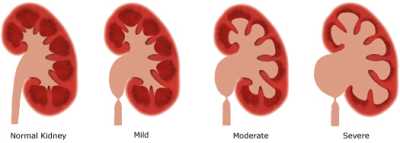
The prognosis for people with hydronephrosis generally depends on how severe the condition is. Mild
cases generally have a better prognosis than moderate and severe conditions. The severity of the
condition is determined by how big the blockage is and how much the kidney has been stretched. In
children with hydronephrosis, the condition may disappear by the first year of life. In fact, in about half of
the cases in which hydronephrosis is detected with an ultrasound before the child is born, the condition
goes away by itself by the time the child is born.
Overall, about 93% of hydronephrosis cases go away without treatment. The cases that persist are
usually those in which the case is more severe. In these cases, the condition usually worsens over time,
kidney function decreases, and surgery is required. Kidney functioning is usually not affected after
surgery.
In children with moderate hydronephrosis, kidney functioning usually does not decrease, growth of the
kidneys remains normal, and the condition usually does not worsen. The kidney usually compensates for
the hydronephrosis in moderate conditions, and often finds a way to function normally. Some children with
moderate hydronephrosis may even get better on their own. In very severe cases of hydronephrosis,
kidney damage may occur.
If a blockage is detected in the unborn child and surgery is used to correct it shortly after birth, kidney
function often improves. Early diagnosis of a blockage that leads to hydronephrosis improves outcome,
because treatment can be implemented earlier. If hydronephrosis is not treated, it can lead to severe
kidney damage and the kidney can wear away.
If both kidneys are affected, kidney failure can result. Kidney failure rarely occurs when one kidney is
affected because the other kidney can usually compensate for the bad one. If the patient only has one
kidney to begin with, however, kidney failure will occur if the condition goes untreated.
In cases of severe kidney abnormalities, the kidneys usually will not function properly regardless of
treatment. In severe cases where an experimental surgery is done while the unborn child is still inside the
mother, the outcome of these children has not yet been improved to date. Follow-up studies in the future
should lead to a better understanding of the prognosis of unborn children that undergo such a surgery.
WHAT ELSE IS HYDRONEPHROSIS KNOWN AS?
Hydronephrosis is also known as pelvocaliectasis and pyeloureterectasis.
WHAT IS THE ORIGIN OF THE TERM, HYDRONEPHROSIS?
Hydronephrosis comes from the Greek word "hydor" meaning "water," the Greek word "nephros" meaning
"kidney," and the Greek word "osis" meaning "condition." Put the words together and you get "water kidney
condition."